Huang Wen-Qing, Lin Qing, Tzeng Chi-Meng
Department of Central Laboratory, Shanghai Children's Hospital, School of Medicine, Shanghai Jiao Tong University, Shanghai, China.
Department of Neurology, The First Affiliated Hospital of Xiamen University, School of Medicine, Xiamen University, Xiamen, Fujian, China.
J Stroke. 2024 May;26(2):131-163. doi: 10.5853/jos.2023.02719. Epub 2024 May 30.
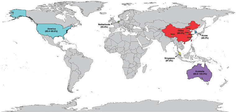
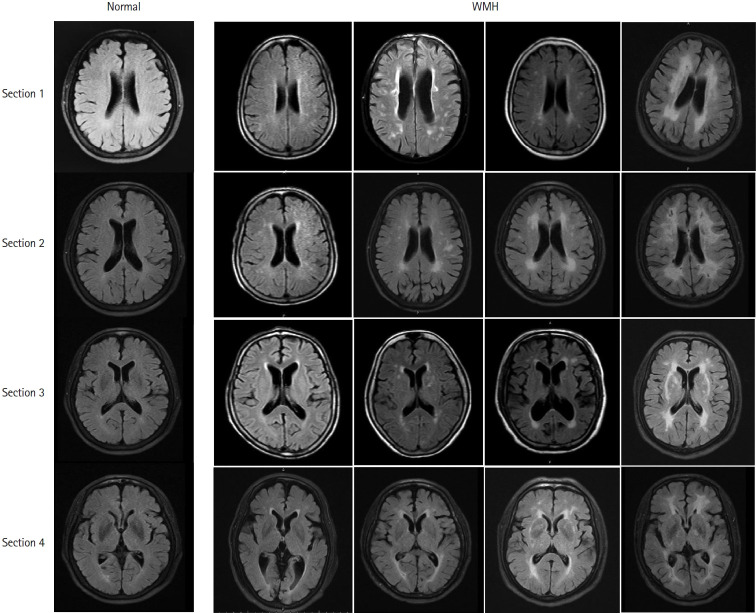
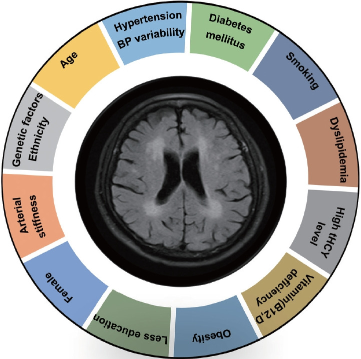
Leukoaraiosis (LA) manifests as cerebral white matter hyperintensities on T2-weighted magnetic resonance imaging scans and corresponds to white matter lesions or abnormalities in brain tissue. Clinically, it is generally detected in the early 40s and is highly prevalent globally in individuals aged >60 years. From the imaging perspective, LA can present as several heterogeneous forms, including punctate and patchy lesions in deep or subcortical white matter; lesions with periventricular caps, a pencil-thin lining, and smooth halo; as well as irregular lesions, which are not always benign. Given its potential of having deleterious effects on normal brain function and the resulting increase in public health burden, considerable effort has been focused on investigating the associations between various risk factors and LA risk, and developing its associated clinical interventions. However, study results have been inconsistent, most likely due to potential differences in study designs, neuroimaging methods, and sample sizes as well as the inherent neuroimaging heterogeneity and multi-factorial nature of LA. In this article, we provided an overview of LA and summarized the current knowledge regarding its epidemiology, neuroimaging classification, pathological characteristics, risk factors, and potential intervention strategies.
脑白质疏松症(LA)在T2加权磁共振成像扫描中表现为脑白质高信号,对应于脑组织中的白质病变或异常。临床上,它通常在40岁出头时被检测到,在全球60岁以上的人群中非常普遍。从影像学角度来看,LA可以呈现多种不同的形式,包括深部或皮质下白质的点状和斑片状病变;伴有脑室周围帽、铅笔细的边缘和光滑晕圈的病变;以及不规则病变,这些病变并不总是良性的。鉴于其对正常脑功能可能产生有害影响以及由此导致的公共卫生负担增加,人们已经投入了大量精力来研究各种风险因素与LA风险之间的关联,并开发其相关的临床干预措施。然而,研究结果并不一致,这很可能是由于研究设计、神经影像学方法、样本量的潜在差异,以及LA固有的神经影像学异质性和多因素性质。在本文中,我们概述了LA,并总结了关于其流行病学、神经影像学分类、病理特征、风险因素和潜在干预策略的当前知识。