Department of Pulmonary and Critical Care Medicine, The First Affiliated Hospital of Soochow University, Suzhou, 215006, China.
Suzhou Key Laboratory for Respiratory Diseases, Suzhou, 215000, China.
Cell Commun Signal. 2024 Jun 6;22(1):313. doi: 10.1186/s12964-024-01639-1.
Non-small-cell lung cancer (NSCLC) accounts for 80-85% of all lung cancer and is the leading cause of cancer-related deaths globally. Although various treatment strategies have been introduced, the 5-year survival rate of patients with NSCLC is only 20-30%. Thus, it remains necessary to study the pathogenesis of NSCLC and develop new therapeutic drugs. Notably, PYK2 has been implicated in the progression of many tumors, including NSCLC, but its detailed mechanism remains unclear. In this study, we aimed to elucidate the mechanisms through which PYK2 promotes NSCLC progression.
The mRNA and protein levels of various molecules were measured using qRT-PCR, western blot (WB), and immunohistochemistry (IHC), respectively. We established stable PYK2 knockdown and overexpression cell lines, and CCK-8, EdU, and clonogenic assays; wound healing, transwell migration, and Matrigel invasion assays; and flow cytometry were employed to assess the phenotypes of tumor cells. Protein interactions were evaluated with co-immunoprecipitation (co-IP), immunofluorescence (IF)-based colocalization, and nucleocytoplasmic separation assays. RNA sequencing was performed to explore the transcriptional regulation mediated by PYK2. Secreted VGF levels were examined using ELISA. Dual-luciferase reporter system was used to detect transcriptional regulation site. PF4618433 (PYK2 inhibitor) and Stattic (STAT3 inhibitor) were used for rescue experiments. A public database was mined to analyze the effect of these molecules on NSCLC prognosis. To investigate the role of PYK2 in vivo, mouse xenograft models of lung carcinoma were established and examined.
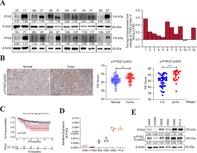
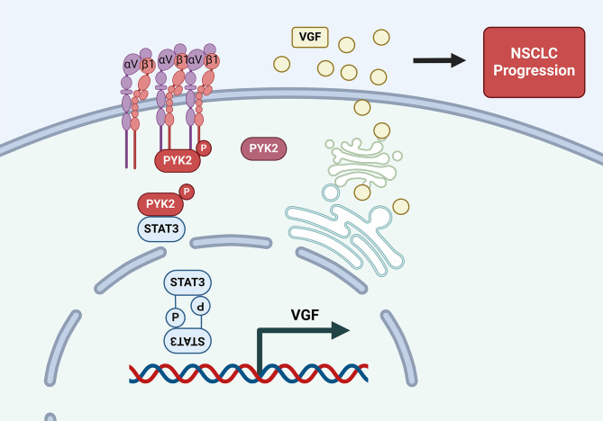
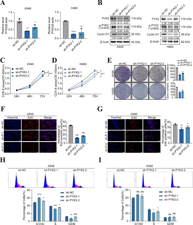
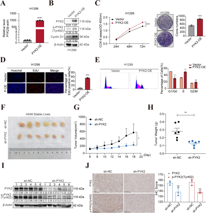
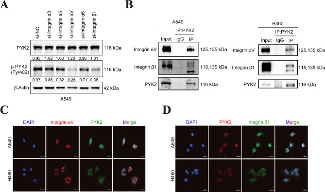
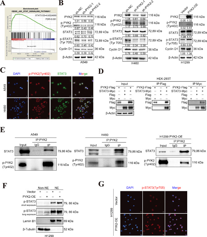
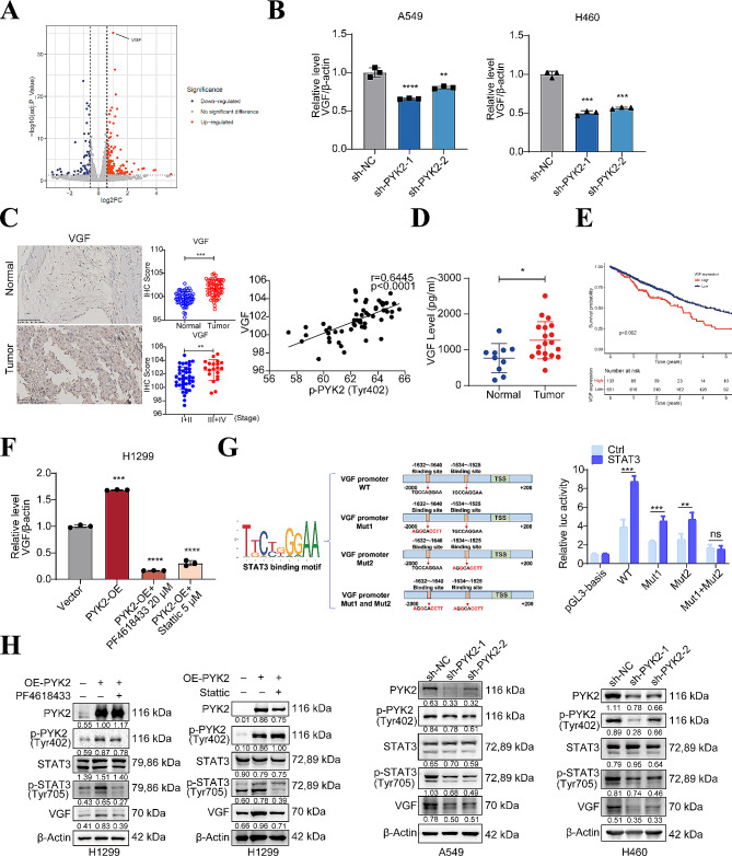
The protein level of PYK2 was higher in human NSCLC tumors than in the adjacent normal tissue, and higher PYK2 expression was associated with poorer prognosis. PYK2 knockdown inhibited the proliferation and motility of tumor cells and caused G1-S arrest and cyclinD1 downregulation in A549 and H460 cells. Meanwhile, PYK2 overexpression had the opposite effect in H1299 cells. The siRNA-induced inhibition of integrins alpha V and beta 1 led to the downregulation of p-PYK2(Tyr402). Activated PYK2 could bind to STAT3 and enhance its phosphorylation at Tyr705, regulating the nuclear accumulation of p-STAT3(Tyr705). This further promoted the expression of VGF, as confirmed by RNA sequencing in a PYK2-overexpressing H1299 cell line and validated by rescue experiments. Two sites in promoter region of VGF gene were confirmed as binding sites of STAT3 by Dual-luciferase assay. Data from the TGCA database showed that VGF was related to the poor prognosis of NSCLC. IHC revealed higher p-PYK2(Tyr402) and VGF expression in lung tumors than in adjacent normal tissues. Moreover, both proteins showed higher levels in advanced TNM stages than earlier ones. A positive linear correlation existed between the IHC score of p-PYK2(Tyr402) and VGF. Knockdown of VGF inhibited tumor progression and reversed the tumor promoting effect of PYK2 overexpression in NSCLC cells. Finally, the mouse model exhibited enhanced tumor growth when PYK2 was overexpressed, while the inhibitors PF4618433 and Stattic could attenuate this effect.
The Integrin αVβ1-PYK2-STAT3-VGF axis promotes NSCLC development, and the PYK2 inhibitor PF4618433 and STAT3 inhibitor Stattic can reverse the pro-tumorigenic effect of high PYK2 expression in mouse models. Our findings provide insights into NSCLC progression and could guide potential therapeutic strategies against NSCLC with high PYK2 expression levels.
非小细胞肺癌(NSCLC)占所有肺癌的 80-85%,是全球癌症相关死亡的主要原因。尽管已经引入了各种治疗策略,但 NSCLC 患者的 5 年生存率仅为 20-30%。因此,仍然有必要研究 NSCLC 的发病机制并开发新的治疗药物。值得注意的是,PYK2 参与了许多肿瘤的进展,包括 NSCLC,但其详细机制仍不清楚。在这项研究中,我们旨在阐明 PYK2 促进 NSCLC 进展的机制。
使用 qRT-PCR、western blot(WB)和免疫组织化学(IHC)分别测量各种分子的 mRNA 和蛋白水平。我们建立了稳定的 PYK2 敲低和过表达细胞系,并进行了 CCK-8、EdU 和集落形成实验;划痕愈合、Transwell 迁移和 Matrigel 侵袭实验;以及流式细胞术来评估肿瘤细胞的表型。使用共免疫沉淀(co-IP)、免疫荧光(IF)共定位和核质分离实验评估蛋白质相互作用。进行 RNA 测序以探索 PYK2 介导的转录调控。使用 ELISA 检测分泌型 VGF 水平。使用双荧光素酶报告系统检测转录调控位点。使用 PYK2 抑制剂 PF4618433 和 STAT3 抑制剂 Stattic 进行挽救实验。挖掘公共数据库分析这些分子对 NSCLC 预后的影响。为了研究 PYK2 在体内的作用,建立并检查了肺腺癌小鼠异种移植模型。
与相邻正常组织相比,人类 NSCLC 肿瘤中的 PYK2 蛋白水平较高,较高的 PYK2 表达与较差的预后相关。PYK2 敲低抑制了肿瘤细胞的增殖和迁移,并导致 A549 和 H460 细胞中的 G1-S 期阻滞和细胞周期蛋白 D1 下调。同时,H1299 细胞中的 PYK2 过表达则产生相反的效果。整合素 alpha V 和 beta 1 的 siRNA 诱导抑制导致 p-PYK2(Tyr402)下调。激活的 PYK2 可以与 STAT3 结合并增强其 Tyr705 磷酸化,调节 p-STAT3(Tyr705)的核积累。这进一步促进了 VGF 的表达,这在 PYK2 过表达的 H1299 细胞系中的 RNA 测序中得到证实,并通过挽救实验得到验证。双荧光素酶测定证实 VGF 基因启动子区域的两个位点是 STAT3 的结合位点。来自 TGCA 数据库的数据表明 VGF 与 NSCLC 的不良预后相关。IHC 显示肺肿瘤中的 p-PYK2(Tyr402)和 VGF 表达高于相邻正常组织。此外,在晚期 TNM 分期中,这两种蛋白的水平高于早期分期。p-PYK2(Tyr402)和 VGF 的 IHC 评分之间存在正线性相关。VGF 的敲低抑制了肿瘤进展,并在 NSCLC 细胞中逆转了 PYK2 过表达的肿瘤促进作用。最后,在 PYK2 过表达的小鼠模型中,肿瘤生长增强,而 PYK2 抑制剂 PF4618433 和 STAT3 抑制剂 Stattic 可以减弱这种作用。
整合素 αVβ1-PYK2-STAT3-VGF 轴促进 NSCLC 的发展,PYK2 抑制剂 PF4618433 和 STAT3 抑制剂 Stattic 可以逆转小鼠模型中高 PYK2 表达的促肿瘤作用。我们的研究结果为 NSCLC 的进展提供了新的见解,并为针对高 PYK2 表达水平的 NSCLC 提供潜在的治疗策略提供了指导。