文献检索文档翻译深度研究
Suppr Zotero 插件Zotero 插件
邀请有礼套餐&价格历史记录

新学期,新优惠

限时优惠:9月1日-9月22日

30天高级会员仅需29元

1天体验卡首发特惠仅需5.99元

了解详情
不再提醒
插件&应用
Suppr Zotero 插件Zotero 插件浏览器插件Mac 客户端Windows 客户端微信小程序
高级版
套餐订阅购买积分包
AI 工具
文献检索文档翻译深度研究
关于我们
关于 Suppr公司介绍联系我们用户协议隐私条款
关注我们

Suppr 超能文献

核心技术专利:CN118964589B侵权必究
粤ICP备2023148730 号-1Suppr @ 2025

鉴定和描述哺乳动物神经嵴细胞上皮到间充质转化和脱层过程中的中间状态。

Identification and characterization of intermediate states in mammalian neural crest cell epithelial to mesenchymal transition and delamination.

机构信息

Stowers Institute for Medical Research, Kansas City, United States.

Department of Anatomy and Cell Biology, University of Kansas Medical Center, Kansas City, United States.

出版信息

Elife. 2024 Jun 14;13:RP92844. doi: 10.7554/eLife.92844.


DOI:10.7554/eLife.92844
PMID:38873887
原文链接:https://pmc.ncbi.nlm.nih.gov/articles/PMC11178358/
Abstract

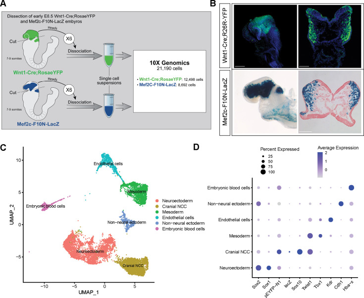
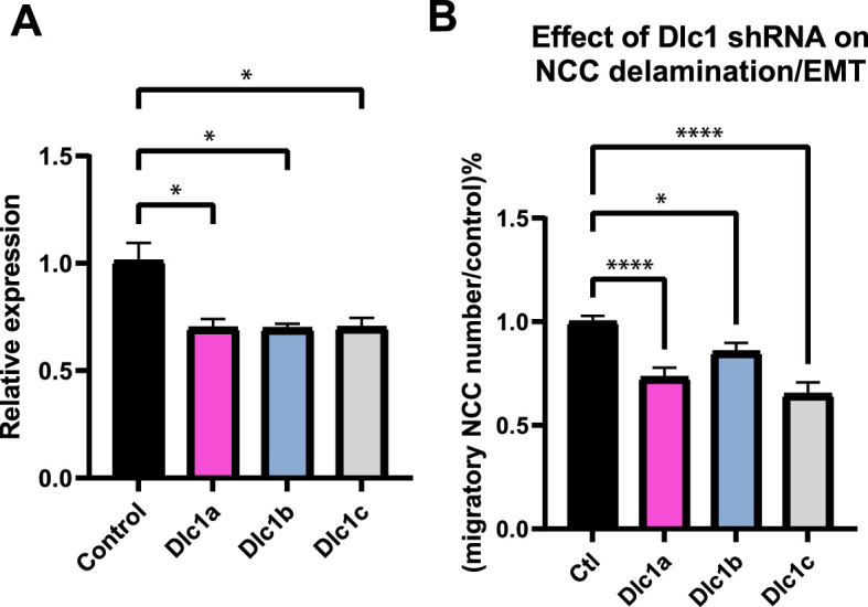
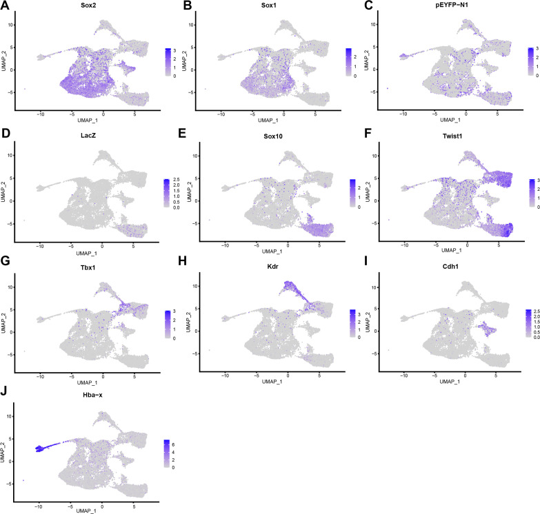
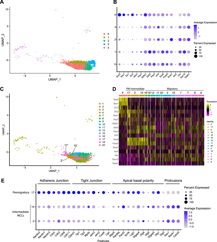
Epithelial to mesenchymal transition (EMT) is a cellular process that converts epithelial cells to mesenchymal cells with migratory potential in developmental and pathological processes. Although originally considered a binary event, EMT in cancer progression involves intermediate states between a fully epithelial and a fully mesenchymal phenotype, which are characterized by distinct combinations of epithelial and mesenchymal markers. This phenomenon has been termed epithelial to mesenchymal plasticity (EMP), however, the intermediate states remain poorly described and it's unclear whether they exist during developmental EMT. Neural crest cells (NCC) are an embryonic progenitor cell population that gives rise to numerous cell types and tissues in vertebrates, and their formation and delamination is a classic example of developmental EMT. However, whether intermediate states also exist during NCC EMT and delamination remains unknown. Through single-cell RNA sequencing of mouse embryos, we identified intermediate NCC states based on their transcriptional signature and then spatially defined their locations in situ in the dorsolateral neuroepithelium. Our results illustrate the importance of cell cycle regulation and functional role for the intermediate stage marker in facilitating mammalian cranial NCC delamination and may provide new insights into mechanisms regulating pathological EMP.

摘要

上皮-间充质转化(EMT)是一种细胞过程,可将上皮细胞转化为具有迁移潜能的间充质细胞,这一过程存在于发育和病理过程中。尽管 EMT 最初被认为是一个二元事件,但在癌症进展过程中,EMT 涉及到完全上皮和完全间充质表型之间的中间状态,这些状态的特征是上皮和间充质标志物的不同组合。这种现象被称为上皮-间充质可塑性(EMP),然而,中间状态仍然描述不足,并且不清楚它们是否存在于发育 EMT 过程中。神经嵴细胞(NCC)是一种胚胎祖细胞群体,可在脊椎动物中产生多种细胞类型和组织,其形成和分离是发育 EMT 的经典范例。然而,NCC EMT 和分离过程中是否也存在中间状态尚不清楚。通过对小鼠胚胎进行单细胞 RNA 测序,我们根据其转录特征确定了中间 NCC 状态,然后在原位空间定义了它们在背外侧神经上皮中的位置。我们的研究结果说明了细胞周期调控的重要性和中间阶段标志物在促进哺乳动物颅神经嵴细胞分离中的功能作用,这可能为调节病理性 EMP 的机制提供新的见解。

https://cdn.ncbi.nlm.nih.gov/pmc/blobs/38f0/11178358/234a70fe0fbd/elife-92844-fig6-figsupp1.jpg
https://cdn.ncbi.nlm.nih.gov/pmc/blobs/38f0/11178358/40f135b8a2df/elife-92844-fig1.jpg
https://cdn.ncbi.nlm.nih.gov/pmc/blobs/38f0/11178358/77f4dd7e15c8/elife-92844-fig1-figsupp1.jpg
https://cdn.ncbi.nlm.nih.gov/pmc/blobs/38f0/11178358/fd0d4b16de7b/elife-92844-fig2.jpg
https://cdn.ncbi.nlm.nih.gov/pmc/blobs/38f0/11178358/3a48bc963e16/elife-92844-fig2-figsupp1.jpg
https://cdn.ncbi.nlm.nih.gov/pmc/blobs/38f0/11178358/165d99e1bdea/elife-92844-fig3.jpg
https://cdn.ncbi.nlm.nih.gov/pmc/blobs/38f0/11178358/a7ce4c2bb3a2/elife-92844-fig4.jpg
https://cdn.ncbi.nlm.nih.gov/pmc/blobs/38f0/11178358/0d11fb481e28/elife-92844-fig4-figsupp1.jpg
https://cdn.ncbi.nlm.nih.gov/pmc/blobs/38f0/11178358/41baf72a27c9/elife-92844-fig5.jpg
https://cdn.ncbi.nlm.nih.gov/pmc/blobs/38f0/11178358/c207beb08886/elife-92844-fig5-figsupp1.jpg
https://cdn.ncbi.nlm.nih.gov/pmc/blobs/38f0/11178358/2dccc66db352/elife-92844-fig5-figsupp2.jpg
https://cdn.ncbi.nlm.nih.gov/pmc/blobs/38f0/11178358/840c30c944dd/elife-92844-fig6.jpg
https://cdn.ncbi.nlm.nih.gov/pmc/blobs/38f0/11178358/234a70fe0fbd/elife-92844-fig6-figsupp1.jpg

相似文献

[1]
Identification and characterization of intermediate states in mammalian neural crest cell epithelial to mesenchymal transition and delamination.

Elife. 2024-6-14

[2]
Identification and characterization of intermediate states in mammalian neural crest cell epithelial to mesenchymal transition and delamination.

bioRxiv. 2024-2-15

[3]
Epithelial to mesenchymal transition during mammalian neural crest cell delamination.

Semin Cell Dev Biol. 2023-3-30

[4]
Cell extrusion - a novel mechanism driving neural crest cell delamination.

bioRxiv. 2024-3-12

[5]
p53 coordinates cranial neural crest cell growth and epithelial-mesenchymal transition/delamination processes.

Development. 2011-3-29

[6]
Prickle1 is required for EMT and migration of zebrafish cranial neural crest.

Dev Biol. 2019-2-2

[7]
Live Imaging of the Dynamics of Mammalian Neural Crest Cell Migration.

Methods Mol Biol. 2022

[8]
Using Xenopus Neural Crest Explants to Study Epithelial-Mesenchymal Transition.

Methods Mol Biol. 2021

[9]
Neural crest cell evolution: how and when did a neural crest cell become a neural crest cell.

Curr Top Dev Biol. 2015-1-20

[10]
Resolving time and space constraints during neural crest formation and delamination.

Curr Top Dev Biol. 2015-1-20

引用本文的文献

[1]
Human ectodermal organoids reveal the cellular origin of DiGeorge Syndrome.

bioRxiv. 2025-8-8

[2]
Regulation and dysregulation of microRNA - transcription factor axes in differentiation and neuroblastoma.

Cell Mol Life Sci. 2025-8-8

[3]
Transition paths across the EMT landscape are dictated by network logic.

Development. 2025-5-21

[4]
Integrated mathematical and experimental modeling uncovers enhanced EMT plasticity upon loss of the DLC1 tumor suppressor.

PLoS Comput Biol. 2025-5-12

[5]
Cell extrusion drives neural crest cell delamination.

Proc Natl Acad Sci U S A. 2025-3-18

[6]
Retinoic acid, an essential component of the roof plate organizer, promotes the spatiotemporal segregation of dorsal neural fates.

Development. 2024-10-1

[7]
Craniofacial chondrogenesis in organoids from human stem cell-derived neural crest cells.

iScience. 2024-3-28

[8]
Cell extrusion - a novel mechanism driving neural crest cell delamination.

bioRxiv. 2024-3-12

本文引用的文献

[1]
Dynamic regulation and requirement for ribosomal RNA transcription during mammalian development.

Proc Natl Acad Sci U S A. 2022-8-2

[2]
Quantitative analysis of nuclear pore complex organization in .

Life Sci Alliance. 2022-7

[3]
Epithelial to mesenchymal transition during mammalian neural crest cell delamination.

Semin Cell Dev Biol. 2023-3-30

[4]
Deficient Mice Recapitulate Craniofacial Phenotype and Reveal Developmental Basis of -Related Frontonasal Dysplasia.

Front Cell Dev Biol. 2022-1-21

[5]
Cellpose: a generalist algorithm for cellular segmentation.

Nat Methods. 2021-1

[6]
Recapitulation of Neural Crest Specification and EMT via Induction from Neural Plate Border-like Cells.

Stem Cell Reports. 2020-9-8

[7]
The development, patterning and evolution of neural crest cell differentiation into cartilage and bone.

Bone. 2020-8

[8]
Guidelines and definitions for research on epithelial-mesenchymal transition.

Nat Rev Mol Cell Biol. 2020-4-16

[9]
Nuclear DLC1 exerts oncogenic function through association with FOXK1 for cooperative activation of MMP9 expression in melanoma.

Oncogene. 2020-3-25

[10]
Integrative Bioinformatics Approaches to Map Potential Novel Genes and Pathways Involved in Ovarian Cancer.

Front Bioeng Biotechnol. 2019-12-17

文献AI研究员

20分钟写一篇综述,助力文献阅读效率提升50倍

立即体验

用中文搜PubMed

大模型驱动的PubMed中文搜索引擎

马上搜索

推荐工具

医学文档翻译智能文献检索