• 文献检索
  • 文档翻译
  • 深度研究
  • 学术资讯
  • Suppr Zotero 插件Zotero 插件
  • 邀请有礼
  • 套餐&价格
  • 历史记录
应用&插件
Suppr Zotero 插件Zotero 插件浏览器插件Mac 客户端Windows 客户端微信小程序
定价
高级版会员购买积分包购买API积分包
服务
文献检索文档翻译深度研究API 文档MCP 服务
关于我们
关于 Suppr公司介绍联系我们用户协议隐私条款
关注我们

Suppr 超能文献

核心技术专利:CN118964589B侵权必究
粤ICP备2023148730 号-1Suppr @ 2026

文献检索

告别复杂PubMed语法,用中文像聊天一样搜索,搜遍4000万医学文献。AI智能推荐,让科研检索更轻松。

立即免费搜索

文件翻译

保留排版,准确专业,支持PDF/Word/PPT等文件格式,支持 12+语言互译。

免费翻译文档

深度研究

AI帮你快速写综述,25分钟生成高质量综述,智能提取关键信息,辅助科研写作。

立即免费体验

中国人眼周围 RNFL 横截面积及其与其他参数的关系。

Peripapillary RNFL cross-sectional area and its association with other parameters in a Chinese population.

机构信息

Beijing Key Laboratory of Intraocular Tumor Diagnosis and Treatment, Beijing Ophthalmology & Visual Sciences Key Lab, Medical Artificial Intelligence Research and Verification Key Laboratory of the Ministry of Industry and Information Technology, Beijing Tongren Eye Center, Beijing Tongren Hospital, Capital Medical University, Beijing, 100730, China.

出版信息

BMC Ophthalmol. 2024 Jun 17;24(1):260. doi: 10.1186/s12886-024-03481-y.

DOI:10.1186/s12886-024-03481-y
PMID:38880871
原文链接:https://pmc.ncbi.nlm.nih.gov/articles/PMC11181622/
Abstract

BACKGROUND

Quantitative analysis of retinal nerve fibers is important for the diagnosis and treatment of optic nerve diseases. Peripapillary retinal nerve fiber layer (RNFL) cross-sectional area may give a more accurate quantitative assessment of retinal nerve fibers than RNFL thickness but there have been no previous reports of the peripapillary RNFL cross-sectional area or other parameters. The purpose of the current study was to determine peripapillary RNFL cross-sectional area and its association with other factors in an adult Chinese population.

METHODS

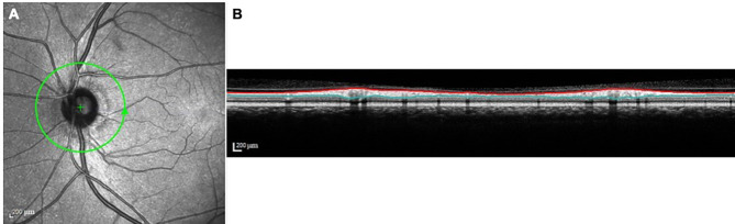
RNFL cross-sectional area was measured during peripapillary circular optical coherence tomography (OCT) scan with a diameter of 12° centered on the optic disc. Correlation between RNFL cross-sectional area and other parameters was evaluated by linear regression analysis in a cross-sectional study of an adult Chinese population.

RESULTS

A total of 2404 eyes from 2404 subjects were examined. Multivariate linear regression analysis showed that larger RNFL cross-sectional area correlated with younger age (p < 0.001), female gender (p = 0.001), no history of diabetes (p = 0.012) and larger optic disc area (p < 0.001).

CONCLUSIONS

Peripapillary RNFL cross-sectional area is correlated positively with optic disc area, suggesting that eyes with larger optic discs have thicker RNFL. Further studies are needed to confirm whether this correlation is due to differences in the numbers of retinal nerve fibers or other factors.

摘要

背景

视网膜神经纤维的定量分析对于视神经疾病的诊断和治疗非常重要。与视网膜神经纤维层(RNFL)厚度相比,视盘周围 RNFL 横截面积可能对视网膜神经纤维提供更准确的定量评估,但目前尚无视盘周围 RNFL 横截面积或其他参数的相关报道。本研究旨在确定成人中国人群的视盘周围 RNFL 横截面积及其与其他因素的关系。

方法

使用直径为 12°的视盘周围环形光相干断层扫描(OCT)测量 RNFL 横截面积,以视盘为中心。在一项成人中国人群的横断面研究中,通过线性回归分析评估 RNFL 横截面积与其他参数之间的相关性。

结果

共检查了 2404 名受试者的 2404 只眼。多变量线性回归分析表明,较大的 RNFL 横截面积与较年轻的年龄(p<0.001)、女性(p=0.001)、无糖尿病史(p=0.012)和较大的视盘面积(p<0.001)相关。

结论

视盘周围 RNFL 横截面积与视盘面积呈正相关,表明视盘较大的眼睛具有较厚的 RNFL。需要进一步的研究来证实这种相关性是由于视网膜神经纤维数量的差异还是其他因素所致。

https://cdn.ncbi.nlm.nih.gov/pmc/blobs/af9e/11181622/33bc27cb1e20/12886_2024_3481_Figa_HTML.jpg

相似文献

1
Peripapillary RNFL cross-sectional area and its association with other parameters in a Chinese population.中国人眼周围 RNFL 横截面积及其与其他参数的关系。
BMC Ophthalmol. 2024 Jun 17;24(1):260. doi: 10.1186/s12886-024-03481-y.
2
Ganglion cell-inner plexiform layer and retinal nerve fiber layer thickness according to myopia and optic disc area: a quantitative and three-dimensional analysis.根据近视和视盘面积分析神经节细胞-内网状层及视网膜神经纤维层厚度:定量与三维分析
BMC Ophthalmol. 2017 Mar 11;17(1):22. doi: 10.1186/s12886-017-0419-1.
3
Optical coherence tomography study of peripapillary retinal nerve fiber layer and choroidal thickness in eyes with tilted optic disc.倾斜视盘眼的视乳头周围视网膜神经纤维层和脉络膜厚度的光学相干断层扫描研究
J Glaucoma. 2015 Jan;24(1):45-50. doi: 10.1097/IJG.0b013e3182883c29.
4
Determinants of normal retinal nerve fiber layer thickness measured by Stratus OCT.通过Stratus OCT测量的正常视网膜神经纤维层厚度的决定因素。
Ophthalmology. 2007 Jun;114(6):1046-52. doi: 10.1016/j.ophtha.2006.08.046. Epub 2007 Jan 8.
5
Optical Coherence Tomography Angiography and Structural Analyses of the Pale Optic Discs: Is It Possible to Differentiate the Cause?光相干断层扫描血管造影和苍白视盘的结构分析:是否可以区分病因?
Curr Eye Res. 2021 Dec;46(12):1876-1885. doi: 10.1080/02713683.2021.1929331. Epub 2021 May 24.
6
Myopic optic disc tilt and the characteristics of peripapillary retinal nerve fiber layer thickness measured by spectral-domain optical coherence tomography.高度近视视盘倾斜与频域光学相干断层扫描测量的视盘周围视网膜神经纤维层厚度的特征。
J Glaucoma. 2012 Apr-May;21(4):260-5. doi: 10.1097/IJG.0b013e31820719e1.
7
Peripapillary retinal nerve fiber layer and optic nerve head characteristics in eyes with situs inversus of the optic disc.视盘反转眼中的视乳头周围视网膜神经纤维层和视神经乳头特征
J Glaucoma. 2015 Apr-May;24(4):306-10. doi: 10.1097/IJG.0b013e31829e1ba2.
8
Usefulness of optical coherence tomography parameters of the optic disc and the retinal nerve fiber layer to differentiate glaucomatous, ocular hypertensive, and normal eyes.视盘和视网膜神经纤维层光学相干断层扫描参数在鉴别青光眼、高眼压症和正常眼方面的应用价值。
J Glaucoma. 2007 Jan;16(1):1-8. doi: 10.1097/01.ijg.0000212215.12180.19.
9
Influence of Myopia on Size of Optic Nerve Head and Retinal Nerve Fiber Layer Thickness Measured by Spectral Domain Optical Coherence Tomography.近视对通过频域光学相干断层扫描测量的视神经乳头大小和视网膜神经纤维层厚度的影响。
Korean J Ophthalmol. 2016 Oct;30(5):335-343. doi: 10.3341/kjo.2016.30.5.335. Epub 2016 Sep 29.
10
Correlation between optic nerve head parameters and retinal nerve fibre layer thickness measured by spectral-domain optical coherence tomography in myopic eyes.近视眼中,应用谱域光学相干断层扫描测量的视神经头参数与视网膜神经纤维层厚度的相关性。
Clin Exp Ophthalmol. 2012 Sep-Oct;40(7):713-20. doi: 10.1111/j.1442-9071.2012.02793.x. Epub 2012 Jun 4.

本文引用的文献

1
High Myopia Normative Database of Peripapillary Retinal Nerve Fiber Layer Thickness to Detect Myopic Glaucoma in a Chinese Population.中国人眼周边视网膜神经纤维层厚度的高度近视正常数据库,用于检测近视性青光眼。
Ophthalmology. 2023 Dec;130(12):1279-1289. doi: 10.1016/j.ophtha.2023.07.022. Epub 2023 Jul 26.
2
[Analysis of the correlation between peripapillary retinal nerve fiber layer cross-sectional area and myopia in individuals aged 50 years and above].[50岁及以上人群视乳头周围视网膜神经纤维层横截面积与近视的相关性分析]
Zhonghua Yan Ke Za Zhi. 2023 Jul 11;59(7):550-556. doi: 10.3760/cma.j.cn112142-20221227-00660.
3
Retinal Nerve Fiber Layer Thickness/Minimum Rim Width Ratio Differentiates Glaucoma From Other Optic Neuropathies.
视网膜神经纤维层厚度/最小视盘边缘宽度比值可区分青光眼与其他视神经病变。
J Glaucoma. 2023 May 1;32(5):435-441. doi: 10.1097/IJG.0000000000002150. Epub 2023 Mar 13.
4
Associated Factors and Distribution of Peripapillary Retinal Nerve Fiber Layer Thickness in Children by Optical Coherence Tomography: A Population-based Study.基于人群的研究:光学相干断层扫描测量儿童视乳头周围视网膜神经纤维层厚度的相关因素及分布。
J Glaucoma. 2022 Aug 1;31(8):666-674. doi: 10.1097/IJG.0000000000002043. Epub 2022 Apr 27.
5
Change of peripapillary retinal nerve fiber layer and choroidal thickness during 4-year myopic progress: Boramae Myopia Cohort Study Report 4.四年近视进展过程中视盘周围视网膜神经纤维层和脉络膜厚度的变化:Boramae 近视队列研究报告 4。
Br J Ophthalmol. 2023 Aug;107(8):1165-1171. doi: 10.1136/bjophthalmol-2021-320596. Epub 2022 Apr 5.
6
Bruch membrane opening-minimum rim width and retinal nerve fiber layer thickness in myopic children.近视儿童的bruch 膜裂孔最小边缘宽度和视网膜神经纤维层厚度。
Photodiagnosis Photodyn Ther. 2021 Dec;36:102524. doi: 10.1016/j.pdpdt.2021.102524. Epub 2021 Sep 3.
7
Change in the ophthalmoscopical optic disc size and shape in a 10-year follow-up: the Beijing Eye Study 2001-2011.10 年随访中眼底视盘大小和形状的变化:北京眼病研究 2001-2011。
Br J Ophthalmol. 2023 Feb;107(2):283-288. doi: 10.1136/bjophthalmol-2021-319632. Epub 2021 Sep 2.
8
Location of Parapapillary Gamma Zone and Vertical Fovea Location. The Beijing Eye Study 2011.视盘旁伽马区和垂直黄斑中心凹位置。2011 年北京眼研究。
Invest Ophthalmol Vis Sci. 2021 Jan 4;62(1):18. doi: 10.1167/iovs.62.1.18.
9
Optic nerve head anatomy in myopia and glaucoma, including parapapillary zones alpha, beta, gamma and delta: Histology and clinical features.近视和青光眼的视神经头解剖结构,包括视盘旁区阿尔法、贝塔、伽马和德尔塔:组织学和临床特征。
Prog Retin Eye Res. 2021 Jul;83:100933. doi: 10.1016/j.preteyeres.2020.100933. Epub 2020 Dec 9.
10
Determinants of Optical Coherence Tomography Parameters in a Population-based Study.一项基于人群的研究中光学相干断层扫描参数的决定因素
Am J Ophthalmol. 2021 Apr;224:163-171. doi: 10.1016/j.ajo.2020.11.015. Epub 2020 Dec 8.