Center for Neurodegeneration and Experimental Therapeutics, University of Alabama at Birmingham, Birmingham, AL 35294, USA.
Center for Cellular and Molecular Imaging, Yale University School of Medicine, New Haven, CT 06510, USA.
Neurobiol Dis. 2024 Sep;199:106595. doi: 10.1016/j.nbd.2024.106595. Epub 2024 Jul 6.
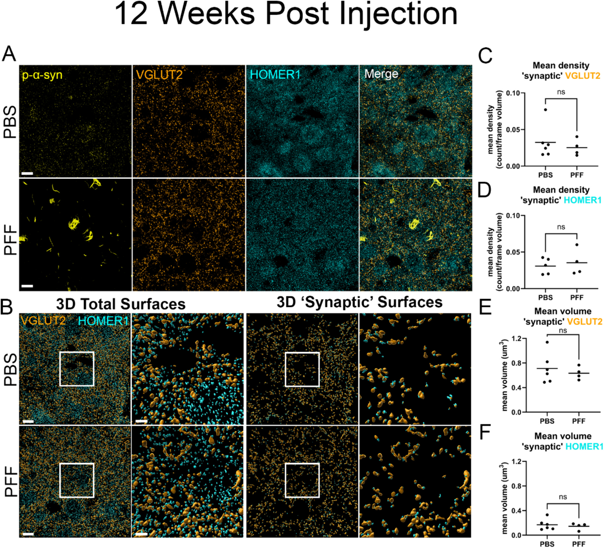
Parkinson's disease (PD) and Dementia with Lewy bodies (DLB) are characterized by neuronal α-synuclein (α-syn) inclusions termed Lewy Pathology, which are abundant in the amygdala. The basolateral amygdala (BLA), in particular, receives projections from the thalamus and cortex. These projections play a role in cognition and emotional processing, behaviors which are impaired in α-synucleinopathies. To understand if and how pathologic α-syn impacts the BLA requires animal models of α-syn aggregation. Injection of α-syn pre-formed fibrils (PFFs) into the striatum induces robust α-syn aggregation in excitatory neurons in the BLA that corresponds with reduced contextual fear conditioning. At early time points after aggregate formation, cortico-amygdala excitatory transmission is abolished. The goal of this project was to determine if α-syn inclusions in the BLA induce synaptic degeneration and/or morphological changes. In this study, we used C57BL/6 J mice injected bilaterally with PFFs in the dorsal striatum to induce α-syn aggregate formation in the BLA. A method was developed using immunofluorescence and three-dimensional reconstruction to analyze excitatory cortico-amygdala and thalamo-amygdala presynaptic terminals closely juxtaposed to postsynaptic densities. The abundance and morphology of synapses were analyzed at 6- or 12-weeks post-injection of PFFs. α-Syn aggregate formation in the BLA did not cause a significant loss of synapses, but cortico-amygdala and thalamo-amygdala presynaptic terminals and postsynaptic densities with aggregates of α-syn show increased volumes, similar to previous findings in human DLB cortex, and in non-human primate models of PD. Transmission electron microscopy showed that asymmetric synapses in mice with PFF-induced α-syn aggregates have reduced synaptic vesicle intervesicular distances, similar to a recent study showing phospho-serine-129 α-syn increases synaptic vesicle clustering. Thus, pathologic α-syn causes major alterations to synaptic architecture in the BLA, potentially contributing to behavioral impairment and amygdala dysfunction observed in synucleinopathies.
帕金森病 (PD) 和路易体痴呆 (DLB) 的特征是神经元α-突触核蛋白 (α-syn) 包含物,称为路易体病理学,其在杏仁核中丰富。特别是基底外侧杏仁核 (BLA),接收来自丘脑和皮层的投射。这些投射在认知和情绪处理中发挥作用,而在α-突触核蛋白病中,这些行为受到损害。为了了解病理性α-syn 是否以及如何影响 BLA,需要有α-syn 聚集的动物模型。将α-syn 预形成纤维 (PFF) 注射到纹状体中会诱导 BLA 中的兴奋性神经元中出现强烈的α-syn 聚集,这与情景性恐惧条件反射的降低相对应。在聚集形成后的早期时间点,皮质-杏仁核兴奋性传递被消除。本项目的目标是确定 BLA 中的α-syn 包含物是否会诱导突触退化和/或形态变化。在这项研究中,我们使用 C57BL/6 J 小鼠将 PFF 双侧注射到背侧纹状体中,以诱导 BLA 中的α-syn 聚集形成。开发了一种使用免疫荧光和三维重建的方法来分析与突触后密度紧密相邻的兴奋性皮质-杏仁核和丘脑-杏仁核前突触末端。在 PFF 注射后 6 或 12 周分析突触的丰度和形态。BLA 中的α-syn 聚集形成不会导致突触明显丢失,但皮质-杏仁核和丘脑-杏仁核前突触末端和与α-syn 聚集物相邻的突触后密度显示出体积增加,类似于先前在人类 DLB 皮层和非人类灵长类动物 PD 模型中的发现。透射电子显微镜显示,在 PFF 诱导的具有α-syn 聚集物的小鼠中,不对称突触的突触小泡间距离减小,类似于最近的一项研究表明磷酸化丝氨酸-129α-syn 增加了突触小泡聚集。因此,病理性α-syn 导致 BLA 中的突触结构发生重大改变,可能导致在突触核蛋白病中观察到的行为障碍和杏仁核功能障碍。