• 文献检索
  • 文档翻译
  • 深度研究
  • 学术资讯
  • Suppr Zotero 插件Zotero 插件
  • 邀请有礼
  • 套餐&价格
  • 历史记录
应用&插件
Suppr Zotero 插件Zotero 插件浏览器插件Mac 客户端Windows 客户端微信小程序
定价
高级版会员购买积分包购买API积分包
服务
文献检索文档翻译深度研究API 文档MCP 服务
关于我们
关于 Suppr公司介绍联系我们用户协议隐私条款
关注我们

Suppr 超能文献

核心技术专利:CN118964589B侵权必究
粤ICP备2023148730 号-1Suppr @ 2026

文献检索

告别复杂PubMed语法,用中文像聊天一样搜索,搜遍4000万医学文献。AI智能推荐,让科研检索更轻松。

立即免费搜索

文件翻译

保留排版,准确专业,支持PDF/Word/PPT等文件格式,支持 12+语言互译。

免费翻译文档

深度研究

AI帮你快速写综述,25分钟生成高质量综述,智能提取关键信息,辅助科研写作。

立即免费体验

掌腱膜纵向纤维和垂直纤维的生化和组织学差异及其创新性的临床意义。

Biochemical and Histological Differences between Longitudinal and Vertical Fibres of Dupuytren's Palmar Aponeurosis and Innovative Clinical Implications.

机构信息

Department of Neurosciences, Institute of Human Anatomy, University of Padova, Via Aristide Gabelli 65, 35127 Padova, Italy.

Plastic and Reconstructive Surgery Unit, University of Padova, Via Nicolò Giustiniani 2, 35128 Padova, Italy.

出版信息

Int J Mol Sci. 2024 Jun 22;25(13):6865. doi: 10.3390/ijms25136865.

DOI:10.3390/ijms25136865
PMID:38999972
原文链接:https://pmc.ncbi.nlm.nih.gov/articles/PMC11241458/
Abstract

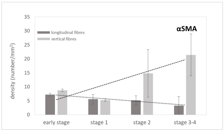
Dupuytren's disease, a chronic and progressive fibroproliferative lesion of the hand, which affects the palmar fascia, has a recurrence rate after selective aponeurotomy of 20-40% at 5 years. This study focused, for the first time, on the microanatomical and histopathological characteristics of the longitudinal and vertical fibres (usually spared during surgery) in the aponeurosis with Dupuytren's disease, in different stages of the Tubiana's classification. Twelve human samples were collected and analysed by immunostaining, Total Collagen Assay, ELISA Immunoassay, and immunoblotting for the Von Willebrand factor, α-Sma, D2-40, CD-68, Total Collagen, Collagen-I and III, IL1β, TNF-α to analyse the blood and lymphatic vascularization, the amount and distribution of collagen, and the inflammation. The results show a progressive increase in the arterial vascularization in the vertical fibres (from 8.8/mm in the early stage to 21.4/mm in stage 3/4), and a parallel progressive decrease in the lymphatic drainage (from 6.2/mm to 2.8/mm), correlated with a local inflammatory context (increase in IL-1β and TNF-α until the stage 2) in both the longitudinal and vertical fibres. The acute inflammation after stage 2 decreased, in favour of a fibrotic action, with the clear synthesis of new collagen (up to ~83 µg/mg), especially Collagen-I. These results clearly demonstrate the involvement of the septa of Legueu and Juvara in the disease pathology and the modifications with the disease's progression. A greater understanding of the pathology becomes fundamental for staging and the adequate therapeutic timing, to obtain the best morpho-functional result and the lowest risk of complications.

摘要

掌腱膜挛缩症是一种手部的慢性、进行性纤维增生性病变,影响掌筋膜,选择性切开术后 5 年的复发率为 20-40%。这项研究首次聚焦于掌腱膜挛缩症患者的腱膜中纵向和垂直纤维(手术中通常保留)的微观解剖和组织病理学特征,这些纤维处于 Tubiana 分类的不同阶段。收集了 12 个人类样本,并通过免疫染色、总胶原测定、ELISA 免疫测定和 Von Willebrand 因子、α-Sma、D2-40、CD-68、总胶原、胶原-I 和 III、IL1β、TNF-α的免疫印迹进行分析,以分析血管和淋巴管的血液和淋巴血管化、胶原的数量和分布,以及炎症。结果表明,垂直纤维中的动脉血管化呈渐进性增加(从早期的 8.8/mm 增加到 3/4 期的 21.4/mm),而淋巴引流呈平行渐进性下降(从 6.2/mm 下降到 2.8/mm),这与纵向和垂直纤维中的局部炎症环境(IL-1β和 TNF-α增加到 2 期)相关。2 期后急性炎症减少,有利于纤维化作用,新胶原(高达约 83µg/mg),特别是胶原-I 的明显合成。这些结果清楚地表明,Legueu 和 Juvara 隔层参与了疾病的病理学改变,并随着疾病的进展而发生改变。更好地了解病理学对于分期和适当的治疗时机至关重要,以获得最佳的形态功能结果和最低的并发症风险。

https://cdn.ncbi.nlm.nih.gov/pmc/blobs/f7c9/11241458/7d3f256518d3/ijms-25-06865-g013.jpg
https://cdn.ncbi.nlm.nih.gov/pmc/blobs/f7c9/11241458/b9d2b38559d8/ijms-25-06865-g001.jpg
https://cdn.ncbi.nlm.nih.gov/pmc/blobs/f7c9/11241458/63007a1b7948/ijms-25-06865-g002.jpg
https://cdn.ncbi.nlm.nih.gov/pmc/blobs/f7c9/11241458/92d322cf944a/ijms-25-06865-g003.jpg
https://cdn.ncbi.nlm.nih.gov/pmc/blobs/f7c9/11241458/f1d8bc4145b2/ijms-25-06865-g004.jpg
https://cdn.ncbi.nlm.nih.gov/pmc/blobs/f7c9/11241458/dc3019fd968b/ijms-25-06865-g005.jpg
https://cdn.ncbi.nlm.nih.gov/pmc/blobs/f7c9/11241458/489dd2637f4a/ijms-25-06865-g006.jpg
https://cdn.ncbi.nlm.nih.gov/pmc/blobs/f7c9/11241458/8d666850a1e9/ijms-25-06865-g007.jpg
https://cdn.ncbi.nlm.nih.gov/pmc/blobs/f7c9/11241458/32cf6803cec9/ijms-25-06865-g008.jpg
https://cdn.ncbi.nlm.nih.gov/pmc/blobs/f7c9/11241458/3b42839b3206/ijms-25-06865-g009.jpg
https://cdn.ncbi.nlm.nih.gov/pmc/blobs/f7c9/11241458/18a27b01bd6d/ijms-25-06865-g010.jpg
https://cdn.ncbi.nlm.nih.gov/pmc/blobs/f7c9/11241458/968279602b9f/ijms-25-06865-g011.jpg
https://cdn.ncbi.nlm.nih.gov/pmc/blobs/f7c9/11241458/89eb38558f02/ijms-25-06865-g012.jpg
https://cdn.ncbi.nlm.nih.gov/pmc/blobs/f7c9/11241458/7d3f256518d3/ijms-25-06865-g013.jpg

相似文献

1
Biochemical and Histological Differences between Longitudinal and Vertical Fibres of Dupuytren's Palmar Aponeurosis and Innovative Clinical Implications.掌腱膜纵向纤维和垂直纤维的生化和组织学差异及其创新性的临床意义。
Int J Mol Sci. 2024 Jun 22;25(13):6865. doi: 10.3390/ijms25136865.
2
Dupuytren's contracture: morphological and biochemical changes in palmar aponeurosis.杜普伊特伦挛缩症:掌腱膜的形态学和生化变化
Hand. 1982 Oct;14(3):237-47. doi: 10.1016/s0072-968x(82)80056-9.
3
Dupuytren's cord involving the septa of Legueu and Juvara: a case report.累及勒格厄和朱瓦拉间隔的杜普伊特伦挛缩索带:一例报告。
J Hand Surg Am. 2002 Mar;27(2):344-6. doi: 10.1053/jhsu.2002.32080.
4
[The fibrous skeleton of the hand : Changes with Dupuytren's contracture].[手部纤维性骨骼:与掌腱膜挛缩症相关的变化]
Orthopade. 2017 Apr;46(4):303-314. doi: 10.1007/s00132-017-3406-6.
5
Biochemical changes in the collagen of the palmar fascia in patients with Dupuytren's disease.掌腱膜挛缩症患者掌腱膜胶原蛋白的生化变化。
J Bone Joint Surg Am. 1981 Jun;63(5):787-97.
6
Fine structural alterations of the palmar aponeurosis in Dupuytren's contracture. A combined scanning and transmission electronmicroscopic examination.掌腱膜在掌腱膜挛缩症中的超微结构改变。扫描电镜与透射电镜联合检查
Zentralbl Allg Pathol. 1988;134(1):15-25.
7
Circulating inflammatory cytokines alter transcriptional activity within fibrotic tissue of Dupuytren's disease patients.循环炎症细胞因子改变了杜普伊特伦挛缩症患者纤维组织内的转录活性。
J Orthop Res. 2022 Mar;40(3):738-749. doi: 10.1002/jor.25059. Epub 2021 May 11.
8
Androgen receptors in Dupuytren's contracture.掌腱膜挛缩症中的雄激素受体。
J Orthop Res. 2002 Jan;20(1):163-8. doi: 10.1016/S0736-0266(01)00072-9.
9
An insight into Dupuytren's contracture.深入了解杜普伊特伦挛缩症。
Ann R Coll Surg Engl. 1992 May;74(3):156-60; discussion 161.
10
Biochemical studies on the collagen of the palmar aponeurosis affected with Dupuytren's disease.对患有杜普伊特伦挛缩症的掌腱膜胶原蛋白的生化研究。
Tohoku J Exp Med. 1984 Apr;142(4):437-43. doi: 10.1620/tjem.142.437.

引用本文的文献

1
Pathological Changes in Extracellular Matrix Composition Orchestrate the Fibrotic Feedback Loop Through Macrophage Activation in Dupuytren's Contracture.掌腱膜挛缩症中细胞外基质成分的病理变化通过巨噬细胞激活来协调纤维化反馈回路。
Int J Mol Sci. 2025 Mar 28;26(7):3146. doi: 10.3390/ijms26073146.
2
The Impact of Sciatic Nerve Injury on Extracellular Matrix of Lower Limb Muscle and Thoracolumbar Fascia: An Observational Study.坐骨神经损伤对下肢肌肉和胸腰筋膜细胞外基质的影响:一项观察性研究。
Int J Mol Sci. 2024 Aug 16;25(16):8945. doi: 10.3390/ijms25168945.

本文引用的文献

1
Targeting Myofibroblasts as a Treatment Modality for Dupuytren Disease.针对肌成纤维细胞的治疗方法在杜普伊特伦挛缩症中的应用
J Hand Surg Am. 2023 Sep;48(9):914-922. doi: 10.1016/j.jhsa.2023.06.007. Epub 2023 Jul 20.
2
Dupuytren's disease: a localised and accessible human fibrotic disorder.杜普伊特伦挛缩症:一种局部且可触及的人类纤维组织疾病。
Trends Mol Med. 2023 Mar;29(3):218-227. doi: 10.1016/j.molmed.2022.12.001. Epub 2022 Dec 23.
3
Dupuytren's Disease-Etiology and Treatment.杜普伊特伦挛缩症-病因与治疗。
Dtsch Arztebl Int. 2021 Nov 19;118(46):781-788. doi: 10.3238/arztebl.m2021.0325.
4
Circulating inflammatory cytokines alter transcriptional activity within fibrotic tissue of Dupuytren's disease patients.循环炎症细胞因子改变了杜普伊特伦挛缩症患者纤维组织内的转录活性。
J Orthop Res. 2022 Mar;40(3):738-749. doi: 10.1002/jor.25059. Epub 2021 May 11.
5
Dupuytren's disease: where do we stand?掌腱膜挛缩症:我们目前的进展如何?
EFORT Open Rev. 2019 Feb 20;4(2):63-69. doi: 10.1302/2058-5241.4.180021. eCollection 2019 Feb.
6
Ponceau S waste: Ponceau S staining for total protein normalization.丽春红 S 废液:用于总蛋白归一化的丽春红 S 染色。
Anal Biochem. 2019 Jun 15;575:44-53. doi: 10.1016/j.ab.2019.03.010. Epub 2019 Mar 23.
7
Recent advances in the understanding of Dupuytren's disease.对掌腱膜挛缩症认识的最新进展。
F1000Res. 2019 Feb 28;8. doi: 10.12688/f1000research.17779.1. eCollection 2019.
8
Assessment of palmar subcutaneous tissue vascularization in patients with Dupuytren's contracture.掌腱膜挛缩症患者手掌皮下组织血管形成的评估
World J Orthop. 2018 Sep 18;9(9):130-137. doi: 10.5312/wjo.v9.i9.130.
9
Hand fasciae innervation: The palmar aponeurosis.手部筋膜的神经支配:掌腱膜。
Clin Anat. 2018 Jul;31(5):677-683. doi: 10.1002/ca.23076. Epub 2018 Apr 14.
10
Dupuytren's Contracture: Current Treatment Methods.杜普伊特伦挛缩症:当前的治疗方法。
Isr Med Assoc J. 2017 Oct;19(10):648-650.