Wani Tariq M, Siddique Ayesha Y, Khan Wajahat N, Rehman Saif, Tram Nguyen K, Tobias Joseph D
Department of Anesthesiology, Sidra Medicine, Doha, Qatar.
Department of Anesthesiology, Hamad General Hospital, Doha, Qatar.
Saudi J Anaesth. 2024 Jul-Sep;18(3):346-351. doi: 10.4103/sja.sja_36_24. Epub 2024 Jun 4.
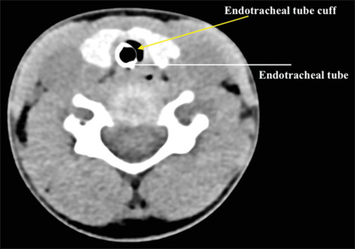
The use of cuffed endotracheal tubes (ETTs) has become the standard of care in pediatric practice. The rationale for the use of a cuffed ETT is to minimize pressure around the cricoid while providing an effective airway seal. However, safe care requires that the cuff lie distal to the cricoid ring following endotracheal intubation. The current study demonstrates the capability of computed tomography (CT) imaging in identifying the position of the cuff of the ETT in intubated patients.
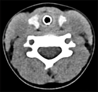
The study included patients ranging in age from 1 month to 10 years who underwent neck and chest CT imaging that required general anesthesia and endotracheal intubation. The location of the ETT and of the cuff within the airway was determined from axial CT images at three levels (proximal, middle, and distal). Anatomical orientations were tabulated, and percent chances of each orientation were determined for the ETT and the cuff.
The study cohort included 42 patients ranging in age from 1 to 114 months. An ETT with a polyvinylchloride cuff was used in 24 patients, and an ETT with a polyurethane cuff was used in 18 patients. The ETT was located near the posterior wall of the trachea in approximately 24-38% of patients, being most likely to be centrally located at the proximal end and at its mid-portion. The middle part of the cuff was most likely to be positioned in the mid-portion of the trachea but tended to skew anteriorly at both the proximal and distal ends.
This is the first study using CT imaging to identify the uniformity of cuff inflation within the trachea in children. With commonly used cuffed ETTs, cuff inflation and the final position of ETT cuff within the tracheal lumen were not uniform. Future investigations are needed to determine the reasons for this asymmetry and its clinical implications.
带套囊气管内插管(ETT)的使用已成为儿科实践中的护理标准。使用带套囊ETT的基本原理是在提供有效气道密封的同时,尽量减少环状软骨周围的压力。然而,安全护理要求气管插管后套囊位于环状软骨环的远端。本研究展示了计算机断层扫描(CT)成像在确定插管患者中ETT套囊位置的能力。
该研究纳入了年龄在1个月至10岁之间,因需要全身麻醉和气管插管而接受颈部和胸部CT成像的患者。从三个层面(近端、中部和远端)的轴向CT图像确定ETT及其套囊在气道内的位置。将解剖方位制成表格,并确定ETT和套囊每种方位的百分比概率。
研究队列包括42名年龄在1至114个月之间的患者。24名患者使用了带聚氯乙烯套囊的ETT,18名患者使用了带聚氨酯套囊的ETT。在大约24% - 38%的患者中,ETT位于气管后壁附近,最有可能位于近端和中部的中心位置。套囊的中部最有可能位于气管中部,但在近端和远端都倾向于向前倾斜。
这是第一项使用CT成像来确定儿童气管内套囊充气均匀性的研究。对于常用的带套囊ETT,套囊充气以及ETT套囊在气管腔内的最终位置并不均匀。需要进一步研究来确定这种不对称的原因及其临床意义。