Dobrynina Larisa A, Kremneva Elena I, Shamtieva Kamila V, Geints Anastasia A, Filatov Alexey S, Gadzhieva Zukhra Sh, Gnedovskaya Elena V, Krotenkova Marina V, Maximov Ivan I
Research Center of Neurology, 125367 Moscow, Russia.
Department of Health and Functioning, Western Norway University of Applied Sciences (HVL), 5063 Bergen, Norway.
Diagnostics (Basel). 2024 Aug 22;14(16):1838. doi: 10.3390/diagnostics14161838.
The cerebral small vessel disease (cSVD) is one of the main causes of vascular and mixed cognitive impairment (CI), and it is associated, in particular, with brain ageing. An understanding of structural tissue changes in an intact cerebral white matter in cSVD might allow one to develop the sensitive biomarkers for early diagnosis and monitoring of disease progression.
to evaluate microstructural changes in the corpus callosum (CC) using diffusion MRI (D-MRI) approaches in cSVD patients with different severity of CI and reveal the most sensitive correlations of diffusion metrics with CI.
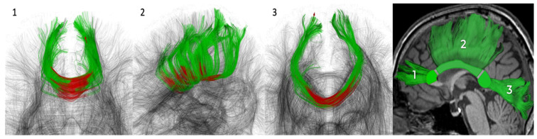
the study included 166 cSVD patients (51.8% women; 60.4 ± 7.6 years) and 44 healthy volunteers (65.9% women; 59.6 ± 6.8 years). All subjects underwent D-MRI (3T) with signal (diffusion tensor and kurtosis) and biophysical (neurite orientation dispersion and density imaging, NODDI, white matter tract integrity, WMTI, multicompartment spherical mean technique, MC-SMT) modeling in three CC segments as well as a neuropsychological assessment.
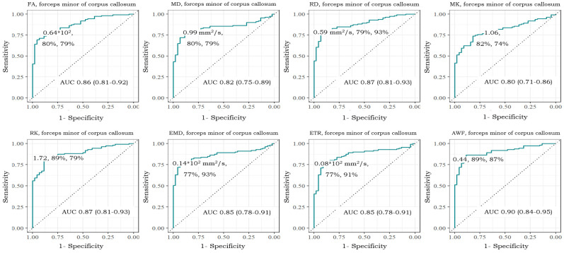
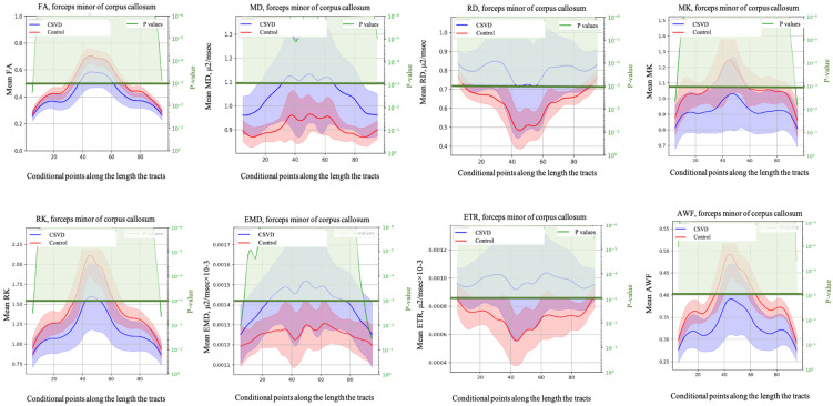
in cSVD patients, microstructural changes were found in all CC segments already at the subjective CI stage, which was found to worsen into mild CI and dementia. More pronounced changes were observed in the forceps minor. Among the signal models FA, MD, MK, RD, and RK, as well as among the biophysical models, MC-SMT (EMD, ETR) and WMTI (AWF) metrics exhibited the largest area under the curve (>0.85), characterizing the loss of microstructural integrity, the severity of potential demyelination, and the proportion of intra-axonal water, respectively. the study reveals the relevance of advanced D-MRI approaches for the assessment of brain tissue changes in cSVD. The identified diffusion biomarkers could be used for the clarification and observation of CI progression.
脑小血管疾病(cSVD)是血管性和混合性认知障碍(CI)的主要原因之一,尤其与脑老化相关。了解cSVD中完整脑白质的结构组织变化可能有助于开发用于疾病早期诊断和监测疾病进展的敏感生物标志物。
使用扩散磁共振成像(D-MRI)方法评估不同CI严重程度的cSVD患者胼胝体(CC)的微观结构变化,并揭示扩散指标与CI最敏感的相关性。
该研究纳入了166例cSVD患者(女性占51.8%;年龄60.4±7.6岁)和44名健康志愿者(女性占65.9%;年龄59.6±6.8岁)。所有受试者均接受了3T的D-MRI检查,对三个CC节段进行了信号(扩散张量和峰度)和生物物理(神经突方向分散和密度成像,NODDI,白质束完整性,WMTI,多室球平均技术,MC-SMT)建模以及神经心理学评估。
在cSVD患者中,在主观CI阶段所有CC节段均发现微观结构变化,且随着病情发展至轻度CI和痴呆而恶化。在小钳中观察到更明显的变化。在信号模型FA、MD、MK、RD和RK以及生物物理模型中,MC-SMT(EMD、ETR)和WMTI(AWF)指标的曲线下面积最大(>0.85),分别表征微观结构完整性丧失、潜在脱髓鞘的严重程度以及轴突内水的比例。该研究揭示了先进的D-MRI方法在评估cSVD脑组织变化方面的相关性。所确定的扩散生物标志物可用于明确和观察CI的进展。