Cruz Gabriel, Jones Daniel T, Lugue Maria T, Heer Manvir, Pace Christopher, Bui Linsey, Silver Scott A
Internal Medicine, Touro University Nevada, Henderson, USA.
Internal Medicine, Touro University California, Vallejo, USA.
Cureus. 2024 Aug 31;16(8):e68305. doi: 10.7759/cureus.68305. eCollection 2024 Aug.
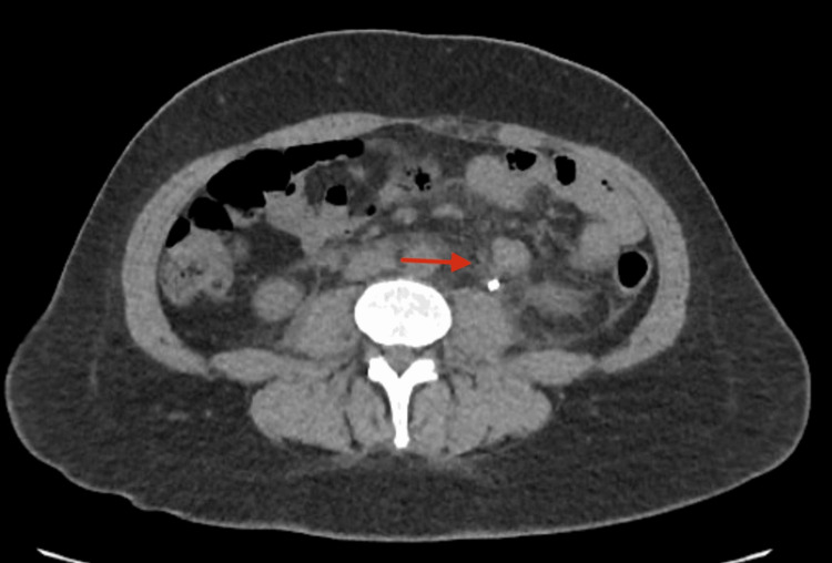
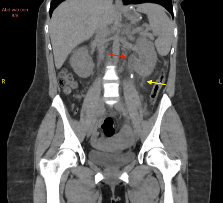
Calyceal rupture, defined as the extravasation of urine from the renal calyces into the perinephric or paranephric spaces, typically results from increased intrapelvic pressure due to urinary tract obstruction. This condition can lead to the formation of a perinephric urinoma and severe complications, such as infection, abscess formation, and impaired renal function. Timely diagnosis and management are crucial to prevent these adverse outcomes. Calyceal rupture often results from urolithiasis, with other causes including strictures, tumors, and congenital abnormalities. The rupture occurs when intrapelvic pressure exceeds the tensile strength of the calyceal walls, leading to urine leakage and potential inflammation or sepsis. Calyceal ruptures are quite rare, with their exact incidence not well-documented due to the infrequency of the condition and potential underreporting. Although relatively uncommon, the condition is more prevalent in individuals with recurrent nephrolithiasis and other predisposing factors. Timely recognition and intervention, guided by imaging studies such as non-contrast CT scans, are essential. Conservative management with medical therapy is effective in many cases, but surgical intervention may be necessary for larger stones or complications. This report presents the case of a 36-year-old female with calyceal rupture secondary to nephrolithiasis, presenting with severe flank pain. Upon initial presentation, the patient underwent a thorough workup, including imaging studies, appropriate medical management, and continuous monitoring. She was stabilized, her pain was effectively managed, and she was discharged with a scheduled outpatient follow-up. This case highlights the importance of early diagnosis, comprehensive management, and vigilant monitoring in preventing complications and promoting favorable outcomes.
肾盏破裂定义为尿液从肾盏渗入肾周或肾旁间隙,通常由尿路梗阻导致肾盂内压力升高引起。这种情况可导致肾周尿瘤形成及严重并发症,如感染、脓肿形成和肾功能损害。及时诊断和处理对于预防这些不良后果至关重要。肾盏破裂常由尿路结石引起,其他原因包括狭窄、肿瘤和先天性异常。当肾盂内压力超过肾盏壁的抗张强度时,就会发生破裂,导致尿液渗漏以及潜在的炎症或脓毒症。肾盏破裂相当罕见,由于这种情况不常见且可能报告不足,其确切发病率尚无充分记录。尽管相对不常见,但在复发性肾结石患者及其他易感因素人群中更为普遍。在诸如非增强CT扫描等影像学检查的指导下及时识别和干预至关重要。在许多情况下,药物保守治疗有效,但对于较大结石或并发症可能需要手术干预。本报告介绍了一例36岁因肾结石继发肾盏破裂的女性病例,患者表现为严重的胁腹疼痛。初诊时,患者接受了全面检查,包括影像学检查、适当的药物治疗和持续监测。她病情稳定,疼痛得到有效控制,出院时安排了门诊随访。该病例强调了早期诊断、综合管理和密切监测对于预防并发症及促进良好预后的重要性。