Department of Nuclear Medicine, University Hospital, LMU Munich, Munich, Germany.
German Center for Neurodegenerative Diseases (DZNE), Munich, Germany.
Mol Neurodegener. 2024 Sep 5;19(1):64. doi: 10.1186/s13024-024-00752-6.
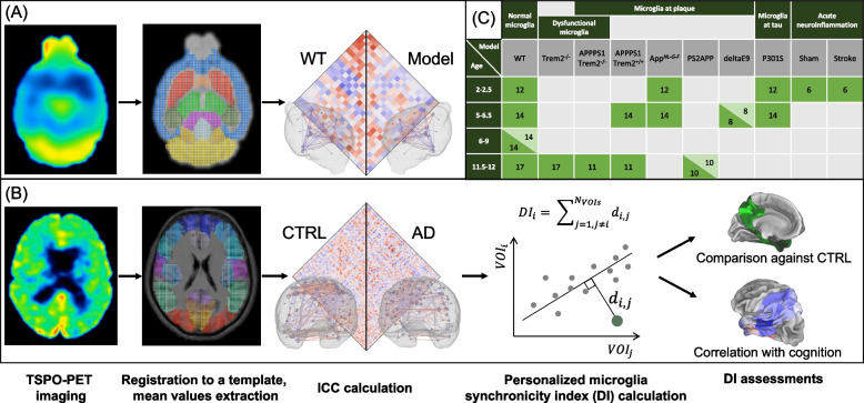
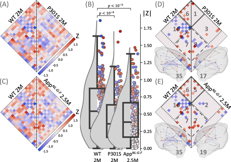
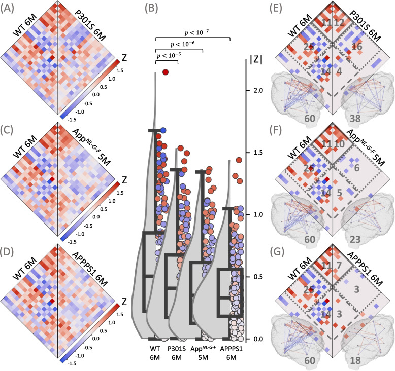
Microglial activation is one hallmark of Alzheimer disease (AD) neuropathology but the impact of the regional interplay of microglia cells in the brain is poorly understood. We hypothesized that microglial activation is regionally synchronized in the healthy brain but experiences regional desynchronization with ongoing neurodegenerative disease. We addressed the existence of a microglia connectome and investigated microglial desynchronization as an AD biomarker.
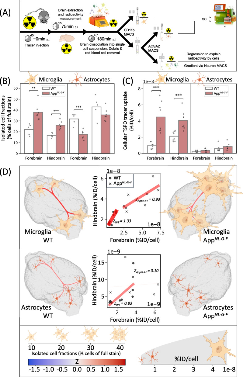
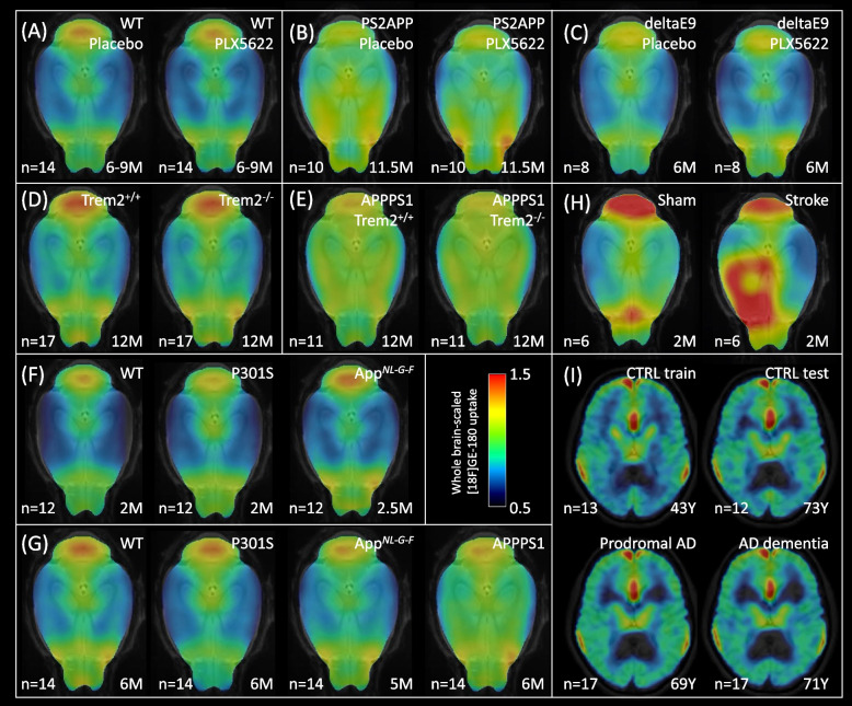
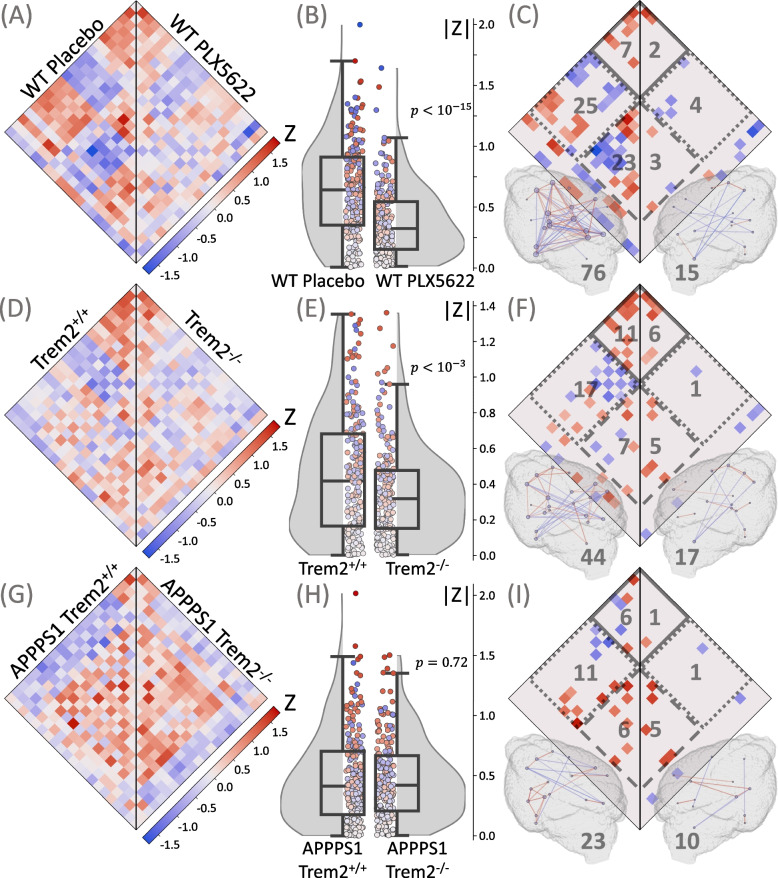
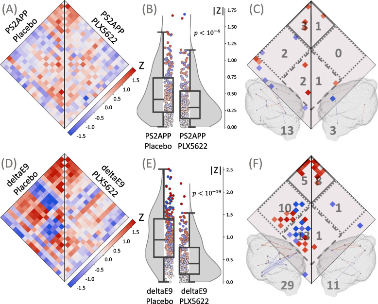
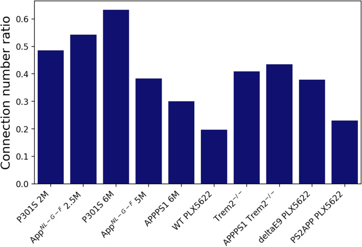
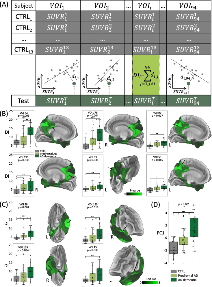
To validate the concept, we performed microglia depletion in mice to test whether interregional correlation coefficients (ICCs) of 18 kDa translocator protein (TSPO)-PET change when microglia are cleared. Next, we evaluated the influence of dysfunctional microglia and AD pathophysiology on TSPO-PET ICCs in the mouse brain, followed by translation to a human AD-continuum dataset. We correlated a personalized microglia desynchronization index with cognitive performance. Finally, we performed single-cell radiotracing (scRadiotracing) in mice to ensure the microglial source of the measured desynchronization.
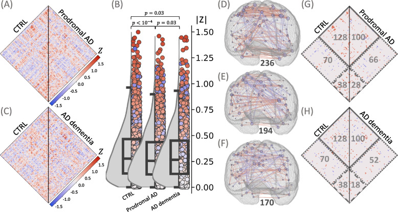
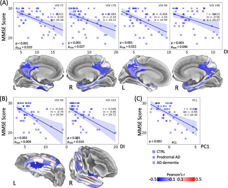
Microglia-depleted mice showed a strong ICC reduction in all brain compartments, indicating microglia-specific desynchronization. AD mouse models demonstrated significant reductions of microglial synchronicity, associated with increasing variability of cellular radiotracer uptake in pathologically altered brain regions. Humans within the AD-continuum indicated a stage-depended reduction of microglia synchronicity associated with cognitive decline. scRadiotracing in mice showed that the increased TSPO signal was attributed to microglia.
Using TSPO-PET imaging of mice with depleted microglia and scRadiotracing in an amyloid model, we provide first evidence that a microglia connectome can be assessed in the mouse brain. Microglia synchronicity is closely associated with cognitive decline in AD and could serve as an independent personalized biomarker for disease progression.
小胶质细胞激活是阿尔茨海默病(AD)神经病理学的一个标志,但大脑中小胶质细胞区域相互作用的影响尚不清楚。我们假设,小胶质细胞在健康大脑中具有区域同步性,但随着进行性神经退行性疾病的发生,会出现区域失同步。我们探讨了小胶质细胞连接组的存在,并研究了小胶质细胞失同步作为 AD 生物标志物的作用。
为了验证这一概念,我们在小鼠中进行小胶质细胞耗竭实验,以测试小胶质细胞清除后,18 kDa 转位蛋白(TSPO)-正电子发射断层扫描(PET)的区域间相关系数(ICCs)是否发生变化。接下来,我们评估了功能失调的小胶质细胞和 AD 病理生理学对小鼠大脑中 TSPO-PET ICC 的影响,然后将其转化为人类 AD 连续体数据集。我们将个性化的小胶质细胞失同步指数与认知表现相关联。最后,我们在小鼠中进行单细胞放射性示踪(scRadiotracing),以确保所测量的失同步源于小胶质细胞。
小胶质细胞耗竭的小鼠在所有脑区均显示出强烈的 ICC 降低,表明小胶质细胞特异性失同步。AD 小鼠模型显示小胶质细胞同步性显著降低,与病理性改变脑区细胞放射性示踪剂摄取的变异性增加有关。AD 连续体中的人类表现出小胶质细胞同步性随认知能力下降而逐渐降低的特征。小鼠的 scRadiotracing 显示,TSPO 信号的增加归因于小胶质细胞。
使用小胶质细胞耗竭的小鼠 TSPO-PET 成像和淀粉样模型中的 scRadiotracing,我们首次提供了在小鼠大脑中评估小胶质细胞连接组的证据。小胶质细胞同步性与 AD 中的认知能力下降密切相关,可能作为疾病进展的独立个性化生物标志物。