Sakamaki Yuri, Shobudani Momoka, Ojiro Ryota, Ozawa Shunsuke, Tang Qian, Zou Xinyu, Ebizuka Yuri, Karasawa Ayumi, Woo Gye-Hyeong, Yoshida Toshinori, Shibutani Makoto
Laboratory of Veterinary Pathology, Division of Animal Life Science, Institute of Agriculture, Tokyo University of Agriculture and Technology, Tokyo, Japan.
Cooperative Division of Veterinary Sciences, Graduate School of Agriculture, Tokyo University of Agriculture and Technology, Tokyo, Japan.
Environ Toxicol. 2025 Jan;40(1):30-53. doi: 10.1002/tox.24413. Epub 2024 Sep 9.
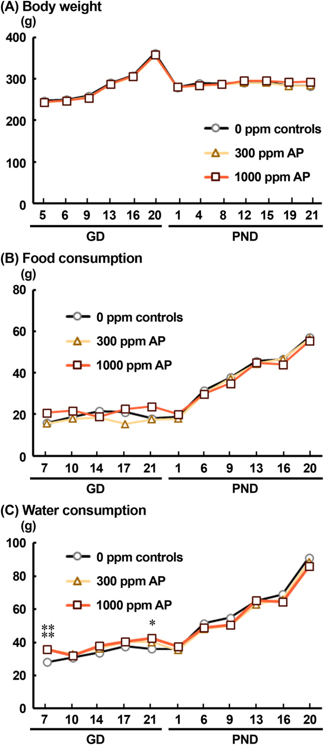
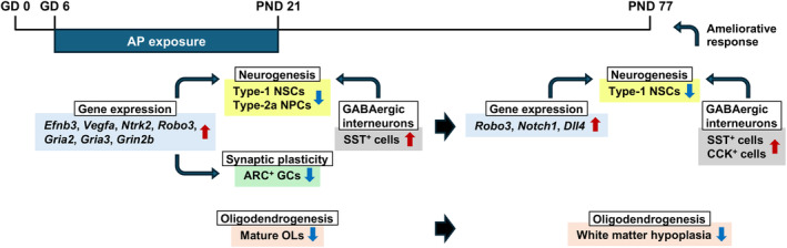
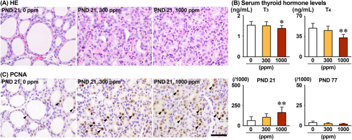
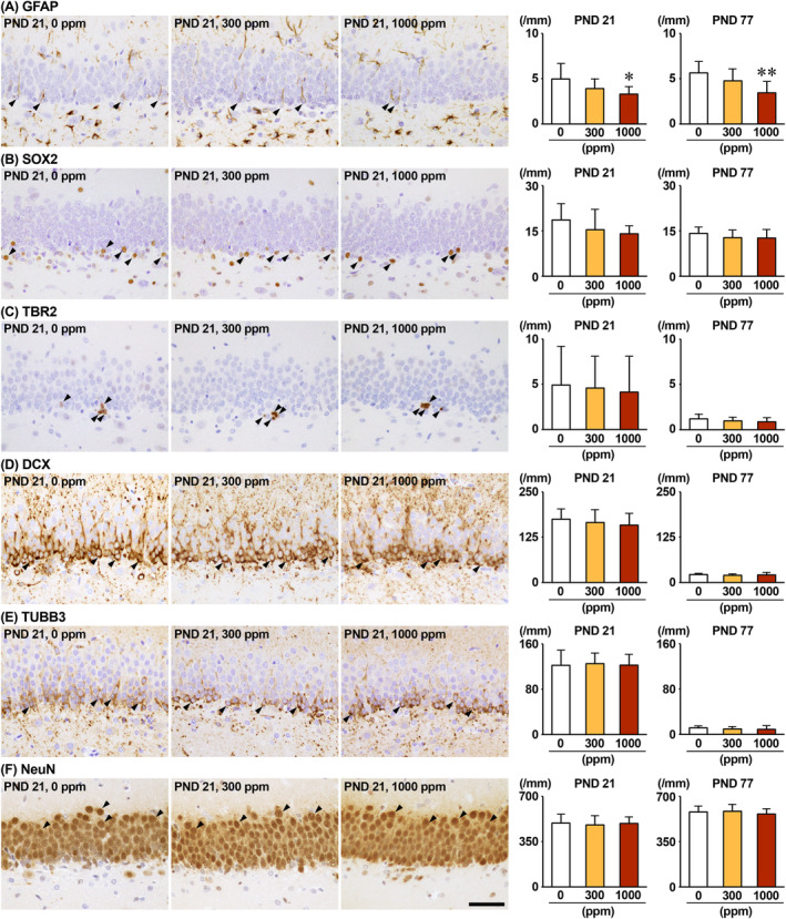
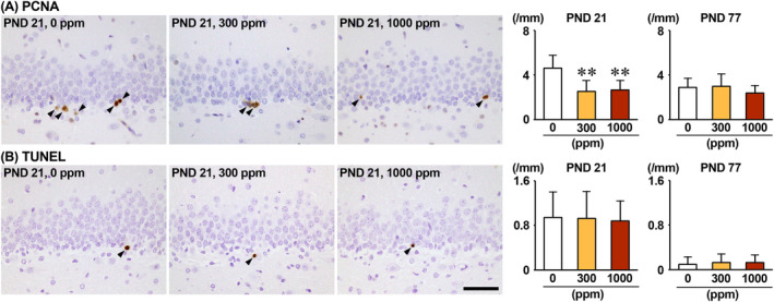
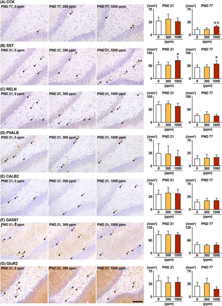
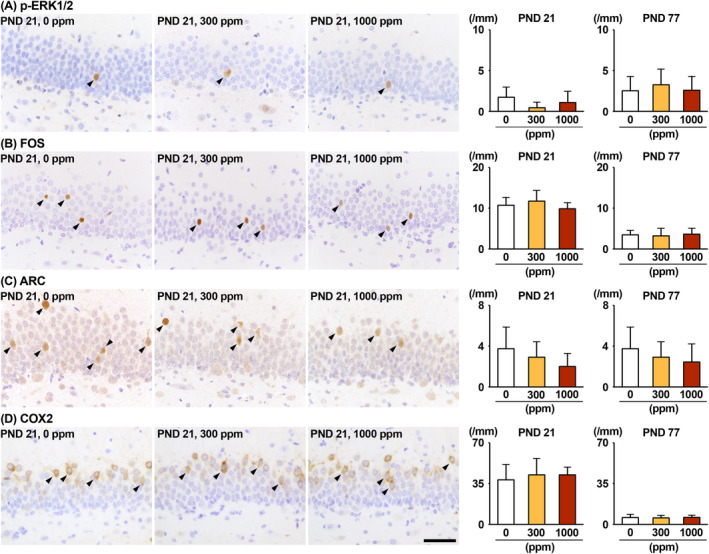
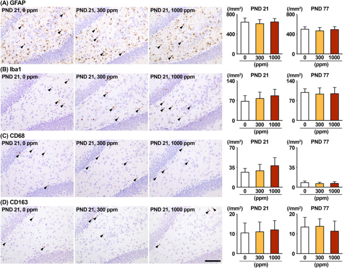
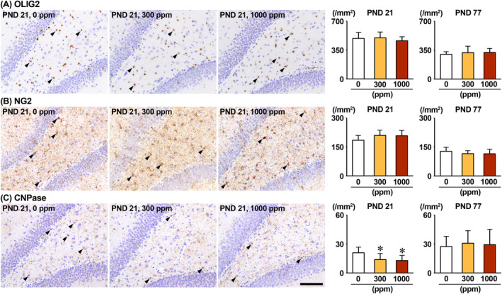
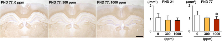
The environmental contaminant perchlorate raises concern for hypothyroidism-related brain disorders in children. This study investigated the effects of developmental perchlorate exposure on hippocampal neurogenesis and oligodendrocyte (OL) development. Pregnant Sprague-Dawley rats were administered with ammonium perchlorate (AP) in drinking water at concentrations of 0 (control), 300, and 1000 ppm from gestation day 6 until weaning [postnatal day (PND) 21]. On PND 21, offspring displayed decreased serum triiodothyronine and thyroxine concentrations at 1000 ppm and thyroid follicular epithelial cell hyperplasia at ≥300 ppm (accompanying increased proliferation activity at 1000 ppm). Hippocampal neurogenesis indicated suppressed proliferation of neurogenic cells at ≥300 ppm, causing decreases in type-1 neural stem cells (NSCs) and type-2a neural progenitor cells. In addition, an increase of SST GABAergic interneurons and decreasing trend for ARC granule cells were observed at 1000 ppm. CNPase mature OLs were decreased in number in the dentate gyrus hilus at ≥300 ppm. At PND 77, thyroid changes had disappeared; however, the decrease of type-1 NSCs and increase of SST interneurons persisted, CCK interneurons were increased, and white matter tissue area was decreased at 1000 ppm. Obtained results suggest an induction of hypothyroidism causing suppressed hippocampal neurogenesis (targeting early neurogenic processes and decreased synaptic plasticity of granule cells involving ameliorative interneuron responses) and suppressed OL maturation during the weaning period. In adulthood, suppression of neurogenesis continued, and white matter hypoplasia was evident. Observed brain changes were similar to those caused by developmental hypothyroidism, suggesting that AP-induced developmental neurotoxicity was due to hypothyroidism.
环境污染物高氯酸盐引发了人们对儿童甲状腺功能减退相关脑部疾病的担忧。本研究调查了发育期暴露于高氯酸盐对海马神经发生和少突胶质细胞(OL)发育的影响。从妊娠第6天到断奶[出生后第(PND)21天],给怀孕的Sprague-Dawley大鼠饮用含高氯酸铵(AP)的水,浓度分别为0(对照)、300和1000 ppm。在PND 21时,1000 ppm组的后代血清三碘甲状腺原氨酸和甲状腺素浓度降低,≥300 ppm组出现甲状腺滤泡上皮细胞增生(1000 ppm组伴随增殖活性增加)。海马神经发生显示,≥300 ppm组神经源性细胞增殖受到抑制,导致1型神经干细胞(NSC)和2a型神经祖细胞减少。此外,在1000 ppm组观察到生长抑素(SST)γ-氨基丁酸能中间神经元增加,弓状核颗粒细胞有减少趋势。≥300 ppm组齿状回门区CNPase成熟OL数量减少。在PND 77时,甲状腺变化已消失;然而,1型NSC减少和SST中间神经元增加的情况仍然存在,胆囊收缩素(CCK)中间神经元增加,1000 ppm组白质组织面积减少。所得结果表明,在断奶期诱发甲状腺功能减退会导致海马神经发生受到抑制(针对早期神经发生过程,降低颗粒细胞的突触可塑性,涉及改善中间神经元反应)以及OL成熟受到抑制。在成年期,神经发生的抑制仍在继续,白质发育不全明显。观察到的脑部变化与发育期甲状腺功能减退引起的变化相似,表明AP诱导的发育神经毒性是由甲状腺功能减退引起的。