Department of Cell and Developmental Biology, Vanderbilt University School of Medicine, Nashville, TN 37232.
Mol Biol Cell. 2024 Nov 1;35(11):br21. doi: 10.1091/mbc.E24-03-0113. Epub 2024 Sep 18.
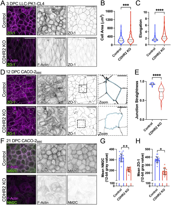
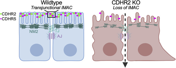
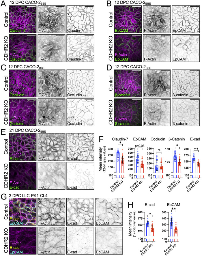
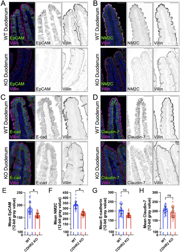
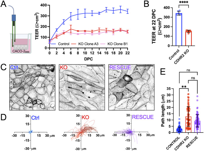
Transporting epithelial cells in the gut and kidney rely on protocadherin-based apical adhesion complexes to organize microvilli that extend into luminal space. In these systems, CDHR2 and CDHR5 localize to the distal ends of microvilli, where they form an intermicrovillar adhesion complex (IMAC) that links the tips of these structures, promotes the formation of a well-ordered array of protrusions, and thus maximizes apical membrane surface area. Recently, we discovered that IMACs can also form between microvilli that extend from neighboring cells, across cell-cell junctions. As an additional point of physical contact between cells, transjunctional IMACs are well positioned to impact the integrity of canonical tight and adherens junctions that form more basolaterally. To begin to test this idea, we examined cell culture and mouse models that lacked CDHR2 expression and were unable to form IMACs. CDHR2 knockout perturbed cell and junction morphology, reduced key components from tight and adherens junctions, impaired barrier function, and increased the motility of single cells within established monolayers. These results support the hypothesis that, in addition to organizing apical microvilli, IMACs provide a layer of cell-cell contact that functions in parallel with canonical tight and adherens junctions to promote epithelial functions.
肠道和肾脏中的上皮细胞运输依赖于原钙黏蛋白基顶膜黏附复合物来组织延伸到腔隙空间的微绒毛。在这些系统中,CDHR2 和 CDHR5 定位于微绒毛的远端,在那里它们形成一个细胞间微绒毛黏附复合物 (IMAC),连接这些结构的尖端,促进突起的有序排列,从而最大限度地增加顶膜表面积。最近,我们发现 IMAC 也可以在从相邻细胞延伸的微绒毛之间形成,穿过细胞-细胞连接处。作为细胞之间的另一个物理接触点,跨连接 IMAC 很好地定位在影响形成更基底外侧的经典紧密和黏附连接的完整性。为了开始验证这个想法,我们检查了缺乏 CDHR2 表达且无法形成 IMAC 的细胞培养和小鼠模型。CDHR2 敲除扰乱了细胞和连接形态,减少了紧密和黏附连接的关键成分,损害了屏障功能,并增加了已建立的单层中单细胞的迁移性。这些结果支持了这样的假设,即除了组织顶膜微绒毛外,IMAC 还提供了一层细胞-细胞接触,与经典紧密和黏附连接平行发挥作用,以促进上皮功能。