Department of Hepatobiliary Surgery and Fujian Institute of Hepatobiliary Surgery, Fujian Medical University Cancer Center, Fujian Medical University Union Hospital, Fuzhou, 350001, Fujian Province, China.
Key Laboratory of The Ministry of Education for Gastrointestinal Cancer, Fujian Medical University, Fuzhou, 350108, Fujian Province, China.
BMC Cancer. 2024 Sep 27;24(1):1191. doi: 10.1186/s12885-024-12817-2.
Identifying primary hepatic angiosarcoma (PHA) preoperatively is challenging, often relying on postoperative pathology. Invasive biopsy increases bleeding risk, emphasizing the importance of early PHA diagnosis through imaging. However, comprehensive summaries of ultrasound, abdominal computed tomography (CT), magnetic resonance imaging (MRI), and whole- body positron emission tomography-CT (PET-CT) in this context are lacking. This study aimed to investigate the comprehensive imaging characteristics of PHA.
Imaging data were collected from 7 patients diagnosed with PHA via pathology between January 2000 and December 2019 in two provincial grade III hospitals. All patients underwent routine color ultrasound examinations before surgery, with 3 patients receiving contrast-enhanced ultrasound (CEUS).CT scans, both plain and enhanced, were performed on 5 patients, and whole-body PET-CT examinations were conducted on 2 patients.
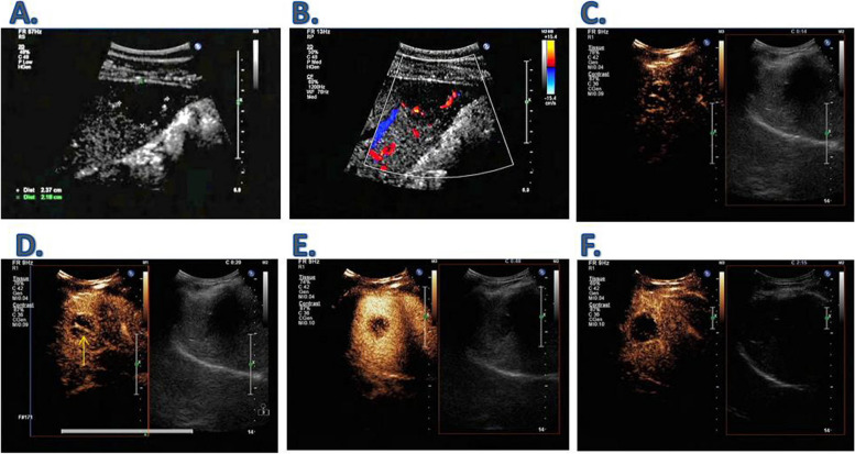
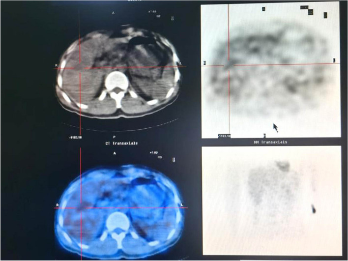
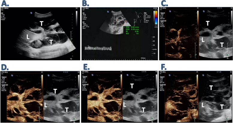
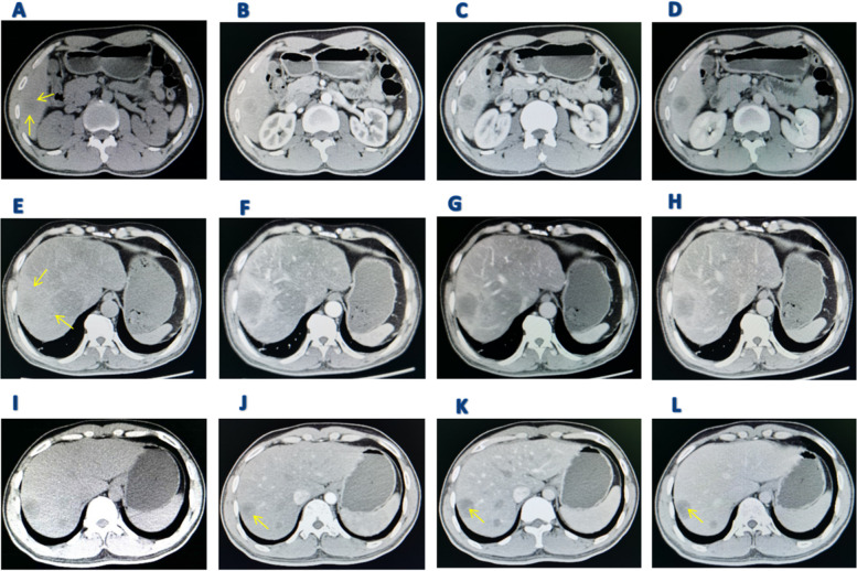
Among the 7 patients with PHA, 4 presented with a single solid intrahepatic mass (2 of which were large), 1 with a single exophytic macroblock type, 1 with a mixed type featuring multiple masses and nodules, and 1 with a multiple nodule type. Conventional ultrasound of PHA showed uneven echoes within the tumor, potentially accompanied by septal zone echoes, and a blood flow grade of 0-I. CEUS displayed early-stage circular high enhancement, a central non-enhancement area, and a "vascular sign" around the tumor. CT scans revealed low-density shadows in the plain scan stage, high peripheral ring enhancement, and punctate nodular enhancement in the arterial phase, with varying intensities and the presence of a "vascular sign." During the portal vein stage, the interior of the tumor was consistently unfilled and exhibited structural disorder. PET-CT showed low-density lesions in the liver and low fluorodeoxyglucose metabolism.
Imaging diagnosis plays a crucial role in PHA diagnosis. When liver tumor imaging matches the above characteristics, consider PHA.
术前识别原发性肝血管肉瘤(PHA)具有挑战性,通常依赖于术后病理。有创性活检会增加出血风险,因此通过影像学进行早期 PHA 诊断尤为重要。然而,目前缺乏关于超声、腹部计算机断层扫描(CT)、磁共振成像(MRI)和全身正电子发射断层扫描-CT(PET-CT)在该背景下的综合总结。本研究旨在探讨 PHA 的全面影像学特征。
收集 2000 年 1 月至 2019 年 12 月期间在两家省级三级医院通过病理诊断为 PHA 的 7 例患者的影像学数据。所有患者均在术前接受常规彩色超声检查,其中 3 例接受了超声造影(CEUS)检查。5 例患者进行了 CT 平扫和增强扫描,2 例患者进行了全身 PET-CT 检查。
在 7 例 PHA 患者中,4 例表现为单个实性肝内肿块(其中 2 个为大肿块),1 例为单个外生巨块型,1 例为混合多个肿块和结节型,1 例为多发结节型。PHA 的常规超声表现为肿瘤内回声不均匀,可能伴有间隔区回声,血流分级为 0-1 级。CEUS 显示早期呈圆形高增强,中央无增强区,肿瘤周围呈“血管征”。CT 扫描显示平扫阶段为低密度阴影,高外周环增强,动脉期呈点状结节增强,增强程度不同,并存在“血管征”。门静脉期时,肿瘤内部始终未填充,结构紊乱。PET-CT 显示肝脏低密度病变和氟脱氧葡萄糖代谢低。
影像学诊断在 PHA 诊断中具有重要作用。当肝肿瘤影像学符合上述特征时,应考虑 PHA。