• 文献检索
  • 文档翻译
  • 深度研究
  • 学术资讯
  • Suppr Zotero 插件Zotero 插件
  • 邀请有礼
  • 套餐&价格
  • 历史记录
应用&插件
Suppr Zotero 插件Zotero 插件浏览器插件Mac 客户端Windows 客户端微信小程序
定价
高级版会员购买积分包购买API积分包
服务
文献检索文档翻译深度研究API 文档MCP 服务
关于我们
关于 Suppr公司介绍联系我们用户协议隐私条款
关注我们

Suppr 超能文献

核心技术专利:CN118964589B侵权必究
粤ICP备2023148730 号-1Suppr @ 2026

文献检索

告别复杂PubMed语法,用中文像聊天一样搜索,搜遍4000万医学文献。AI智能推荐,让科研检索更轻松。

立即免费搜索

文件翻译

保留排版,准确专业,支持PDF/Word/PPT等文件格式,支持 12+语言互译。

免费翻译文档

深度研究

AI帮你快速写综述,25分钟生成高质量综述,智能提取关键信息,辅助科研写作。

立即免费体验

自然杀伤 T 细胞过度激活加重支气管肺发育不良小鼠的肺损伤。

Over-activation of iNKT cells aggravate lung injury in bronchopulmonary dysplasia mice.

机构信息

Department of Pediatrics, The Affiliated Hospital of Jiangsu University, 438 Jiefang Road, Zhenjiang, 212001, China.

International Genome Center, Jiangsu University, 301 Xuefu Road, Zhenjiang, 212013, China.

出版信息

Redox Biol. 2024 Nov;77:103370. doi: 10.1016/j.redox.2024.103370. Epub 2024 Sep 24.

DOI:10.1016/j.redox.2024.103370
PMID:39342744
原文链接:https://pmc.ncbi.nlm.nih.gov/articles/PMC11470607/
Abstract

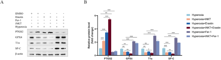
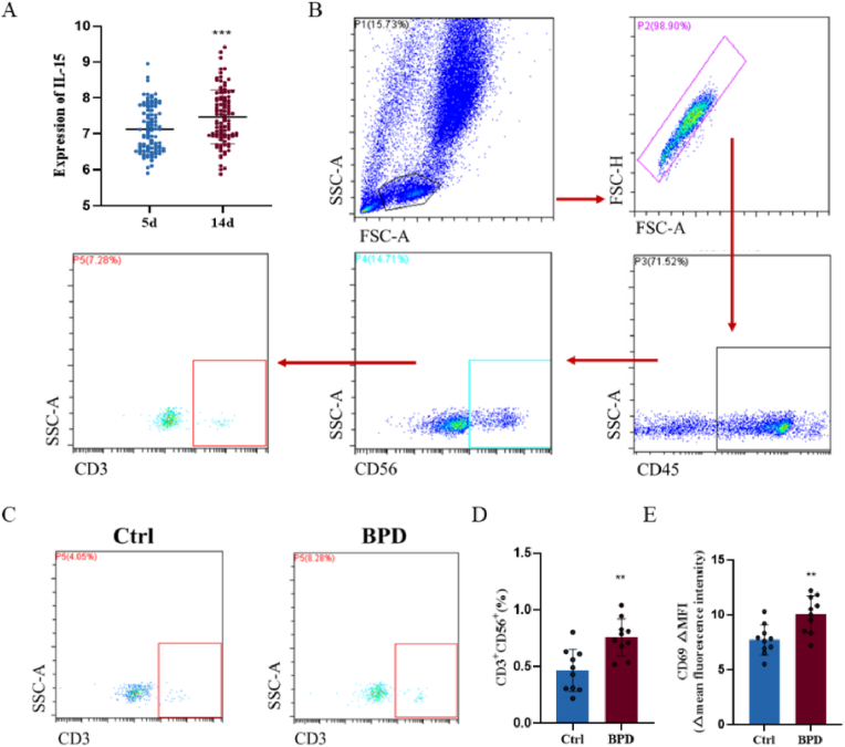
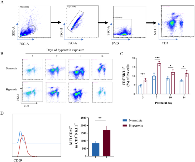
Bronchopulmonary dysplasia (BPD) is a severe lung disease in preterm infants, the abnormal proliferate and differentiate ability of type II epithelial cells (AEC II) is the key to the pathological basis of BPD. Mechanisms regarding abnormal AEC II in BPD remain unclear. The present work investigated the role and mechanisms of invariant natural killer T (iNKT) cells in lung disorder in BPD using public datasets, clinical samples, a hyperoxia-induced BPD mouse model and AEC II-iNKT cells transwell co-culture system. Firstly, we found that the NKT cells development factor IL-15 increased over time in patients with BPD in public databases, and clinically collected peripheral blood NKT cells in patients with BPD were increased. Subsequently, the percentage of iNKT cells increased in hyperoxia group compared with normoxia group, with the highest at P7, accompanied by increased activation with abnormal lung development. The administration of anti-CD1d neutralizing antibody to inhibit iNKT cells could alleviate the abnormal lung development of hyperoxia group mice, while α-GalCer administration could aggravate lung injury in hyperoxia group mice, and adoptive transfer of iNKT cells could aggravate the abnormal lung development in hyperoxia group mice. In addition, to further verify the role of iNKT cells on AEC II, AEC II-iNKT cells co-culture system was established. The presence of iNKT cells could aggravate the abnormal expression of SP-C and T1α under hyperoxia. Meanwhile, RNA-seq analysis showed that ferroptosis-related genes were highly expressed in AEC II co-cultured with iNKT cells under hyperoxia. We further validated the effect of the presence of iNKT cells under hyperoxia environment on AEC II ferroptosis levels, suggested that iNKT cells promote AEC II ferroptosis under hyperoxia, accompanied by decreased expression of SP-C and T1α. Our study found that the recruitment of iNKT cells in the lung may be an important cause of alveolarization disorder in BPD.

摘要

支气管肺发育不良(BPD)是一种严重的早产儿肺部疾病,II 型上皮细胞(AEC II)异常增殖和分化能力是 BPD 病理基础的关键。BPD 中异常 AEC II 的发生机制尚不清楚。本研究利用公共数据集、临床样本、高氧诱导的 BPD 小鼠模型和 AEC II-iNKT 细胞 Transwell 共培养系统,探讨了固有自然杀伤 T(iNKT)细胞在 BPD 肺部疾病中的作用和机制。首先,我们在公共数据库中发现,BPD 患者的 NKT 细胞发育因子 IL-15 随时间推移而增加,且临床采集的 BPD 患者外周血 NKT 细胞也增加。随后,与常氧组相比,高氧组中 iNKT 细胞的比例增加,在 P7 时达到最高,同时伴有异常肺发育的激活。用抗 CD1d 中和抗体抑制 iNKT 细胞可减轻高氧组小鼠的异常肺发育,而 α-GalCer 给药可加重高氧组小鼠的肺损伤,过继转移 iNKT 细胞可加重高氧组小鼠的异常肺发育。此外,为了进一步验证 iNKT 细胞对 AEC II 的作用,建立了 AEC II-iNKT 细胞共培养系统。在高氧条件下,iNKT 细胞的存在可加重 SP-C 和 T1α 的异常表达。同时,RNA-seq 分析显示,在高氧共培养条件下,AEC II 中与铁死亡相关的基因高度表达。我们进一步验证了在高氧环境下 iNKT 细胞存在对 AEC II 铁死亡水平的影响,表明 iNKT 细胞在高氧条件下促进 AEC II 铁死亡,同时 SP-C 和 T1α 的表达降低。本研究发现,肺内 iNKT 细胞的募集可能是 BPD 肺泡化障碍的重要原因。

https://cdn.ncbi.nlm.nih.gov/pmc/blobs/695c/11470607/2ecf1255eaf1/gr8.jpg
https://cdn.ncbi.nlm.nih.gov/pmc/blobs/695c/11470607/a9cd5bf43a06/ga1.jpg
https://cdn.ncbi.nlm.nih.gov/pmc/blobs/695c/11470607/1b7c90d0b805/gr1.jpg
https://cdn.ncbi.nlm.nih.gov/pmc/blobs/695c/11470607/99b7caf48a6a/gr2.jpg
https://cdn.ncbi.nlm.nih.gov/pmc/blobs/695c/11470607/b718c77255d2/gr3.jpg
https://cdn.ncbi.nlm.nih.gov/pmc/blobs/695c/11470607/09690283152c/gr4.jpg
https://cdn.ncbi.nlm.nih.gov/pmc/blobs/695c/11470607/1d57845f8343/gr5.jpg
https://cdn.ncbi.nlm.nih.gov/pmc/blobs/695c/11470607/97169759a588/gr6.jpg
https://cdn.ncbi.nlm.nih.gov/pmc/blobs/695c/11470607/7496a5c617ff/gr7.jpg
https://cdn.ncbi.nlm.nih.gov/pmc/blobs/695c/11470607/2ecf1255eaf1/gr8.jpg

相似文献

1
Over-activation of iNKT cells aggravate lung injury in bronchopulmonary dysplasia mice.自然杀伤 T 细胞过度激活加重支气管肺发育不良小鼠的肺损伤。
Redox Biol. 2024 Nov;77:103370. doi: 10.1016/j.redox.2024.103370. Epub 2024 Sep 24.
2
[Role and mechanism of epithelial-mesenchymal transition in a rat model of bronchopulmonary dysplasia induced by hyperoxia exposure].[高氧暴露诱导的支气管肺发育不良大鼠模型中上皮-间质转化的作用及机制]
Zhongguo Dang Dai Er Ke Za Zhi. 2024 Jul 15;26(7):765-773. doi: 10.7499/j.issn.1008-8830.2312112.
3
Recombinant CXCL17 Treatment Alleviates Hyperoxia-Induced Lung Apoptosis and Inflammation In Vivo and Vitro by Activating the AKT Pathway: A Possible Therapeutic Approach for Bronchopulmonary Dysplasia.重组 CXCL17 治疗通过激活 AKT 通路缓解体内和体外高氧诱导的肺细胞凋亡和炎症:一种支气管肺发育不良的潜在治疗方法。
Mol Biotechnol. 2024 Sep;66(9):2349-2361. doi: 10.1007/s12033-023-00866-0. Epub 2023 Sep 14.
4
Interleukin-11 Is Involved in Hyperoxia-induced Bronchopulmonary Dysplasia in Newborn Mice by Mediating Epithelium-Fibroblast Cross-talk.白细胞介素-11通过介导上皮-成纤维细胞相互作用参与新生小鼠高氧诱导的支气管肺发育不良。
Inflammation. 2025 Apr;48(2):796-805. doi: 10.1007/s10753-024-02089-0. Epub 2024 Jul 24.
5
CD11b(+) Mononuclear Cells Mitigate Hyperoxia-Induced Lung Injury in Neonatal Mice.CD11b(+)单核细胞减轻新生小鼠高氧诱导的肺损伤。
Am J Respir Cell Mol Biol. 2016 Feb;54(2):273-83. doi: 10.1165/rcmb.2014-0395OC.
6
ILC2 regulates hyperoxia-induced lung injury via an enhanced Th17 cell response in the BPD mouse model.ILC2 通过增强 BPD 小鼠模型中的 Th17 细胞反应来调节高氧诱导的肺损伤。
BMC Pulm Med. 2023 May 30;23(1):188. doi: 10.1186/s12890-023-02474-9.
7
Recruitment of PVT1 Enhances YTHDC1-Mediated m6A Modification of IL-33 in Hyperoxia-Induced Lung Injury During Bronchopulmonary Dysplasia.招募 PVT1 增强了 YTHDC1 介导的 IL-33 在支气管肺发育不良期间高氧诱导的肺损伤中的 m6A 修饰。
Inflammation. 2024 Apr;47(2):469-482. doi: 10.1007/s10753-023-01923-1. Epub 2023 Nov 2.
8
Loss of microRNA-30a and sex-specific effects on the neonatal hyperoxic lung injury.miR-30a 的缺失及其对新生鼠高氧肺损伤的性别特异性影响。
Biol Sex Differ. 2023 Aug 8;14(1):50. doi: 10.1186/s13293-023-00535-6.
9
Impaired Autophagic Activity Contributes to the Pathogenesis of Bronchopulmonary Dysplasia. Evidence from Murine and Baboon Models.自噬活性受损导致支气管肺发育不良的发病机制。来自小鼠和狒狒模型的证据。
Am J Respir Cell Mol Biol. 2020 Sep;63(3):338-348. doi: 10.1165/rcmb.2019-0445OC.
10
The Role of Sphingolipid Signaling in Oxidative Lung Injury and Pathogenesis of Bronchopulmonary Dysplasia.鞘脂信号在氧化肺损伤和支气管肺发育不良发病机制中的作用。
Int J Mol Sci. 2022 Jan 23;23(3):1254. doi: 10.3390/ijms23031254.

引用本文的文献

1
Investigating mitophagy mechanisms in bronchopulmonary dysplasia through bioinformatics.通过生物信息学研究支气管肺发育不良中的线粒体自噬机制。
Pediatr Res. 2025 Aug 9. doi: 10.1038/s41390-025-04319-z.
2
The tryptophan metabolite 3-hydroxyanthranilic acid alleviates hyperoxia-induced bronchopulmonary dysplasia via inhibiting ferroptosis.色氨酸代谢产物3-羟基邻氨基苯甲酸通过抑制铁死亡减轻高氧诱导的支气管肺发育不良。
Redox Biol. 2025 May;82:103579. doi: 10.1016/j.redox.2025.103579. Epub 2025 Mar 8.
3
Safety, Efficacy and Bio-Distribution Analysis of Exosomes Derived From Human Umbilical Cord Mesenchymal Stem Cells for Effective Treatment of Bronchopulmonary Dysplasia by Intranasal Administration in Mice Model.
人脐带间充质干细胞来源的外泌体经鼻给药对小鼠模型支气管肺发育不良有效治疗的安全性、有效性及生物分布分析
Int J Nanomedicine. 2025 Feb 27;20:2521-2553. doi: 10.2147/IJN.S501843. eCollection 2025.