• 文献检索
  • 文档翻译
  • 深度研究
  • 学术资讯
  • Suppr Zotero 插件Zotero 插件
  • 邀请有礼
  • 套餐&价格
  • 历史记录
应用&插件
Suppr Zotero 插件Zotero 插件浏览器插件Mac 客户端Windows 客户端微信小程序
定价
高级版会员购买积分包购买API积分包
服务
文献检索文档翻译深度研究API 文档MCP 服务
关于我们
关于 Suppr公司介绍联系我们用户协议隐私条款
关注我们

Suppr 超能文献

核心技术专利:CN118964589B侵权必究
粤ICP备2023148730 号-1Suppr @ 2026

文献检索

告别复杂PubMed语法,用中文像聊天一样搜索,搜遍4000万医学文献。AI智能推荐,让科研检索更轻松。

立即免费搜索

文件翻译

保留排版,准确专业,支持PDF/Word/PPT等文件格式,支持 12+语言互译。

免费翻译文档

深度研究

AI帮你快速写综述,25分钟生成高质量综述,智能提取关键信息,辅助科研写作。

立即免费体验

Scheimpflug 成像与其他筛查指数在诊断圆锥角膜和圆锥角膜疑似患者中的比较。

Comparison of a Scheimpflug imaging with other screening indices in diagnosing keratoconus and keratoconus suspect.

机构信息

School of Medicine, Nankai University, Tianjin, China.

Tianjin Key Lab of Ophthalmology and Visual Science, Tianjin Eye Institute, Tianjin Eye Hospital, Nankai University Affiliated Eye Hospital, Tianjin, China.

出版信息

Sci Rep. 2024 Oct 5;14(1):23187. doi: 10.1038/s41598-024-74497-z.

DOI:10.1038/s41598-024-74497-z
PMID:39369097
原文链接:https://pmc.ncbi.nlm.nih.gov/articles/PMC11455890/
Abstract

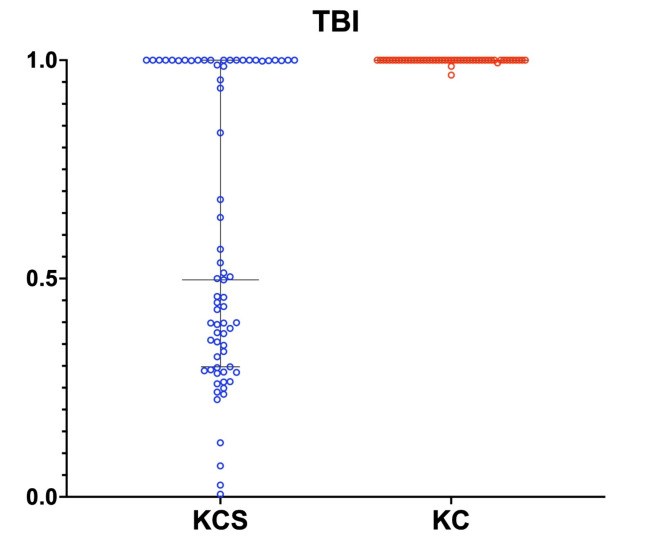
Keratoconus (KC) is an irreversible blinding eye disease; therefore, early screening of KC suspects (KCS) is crucial for protecting patients' quality of life. Scheimpflug imaging is a commonly used screening device in clinical practice. We aimed to evaluate the diagnostic ability of a Scheimpflug imaging device (Scansys) for KC and KCS and compared it with other Scheimpflug-based devices (Pentacam and Corvis ST). This prospective case-control study included 107 normal eyes, 72 KCS, and 57 KC. Scansys screening index Keratoconus probability (KCP) showed excellent performance in diagnosing KC at a cutoff value of 16.4 (area under the receiver operating characteristic [AUROC] = 1.000), with 100% sensitivity and 98.11% specificity. KCP had a better KCS diagnostic ability at a cutoff value of 8.9 (AUROC = 0.813) than Corvis biomechanical index (CBI, AUROC = 0.764), reaching 67.61% sensitivity and 85.85% specificity. Pentacam screening index Belin/Ambrósio enhanced ectasia display deviation (BAD-D) showed the best performance with 92.96% sensitivity and 89.62% specificity at a cutoff value of 1.525 (AUROC = 0.970) in diagnosing KCS. Scansys provides accurate KCP parameters in diagnosing KC; however, the efficiency of diagnosing KCS should be further optimized.

摘要

圆锥角膜(KC)是一种不可逆转的致盲眼病;因此,对 KC 疑似患者(KCS)进行早期筛查对于保护患者的生活质量至关重要。Scheimpflug 成像仪是临床实践中常用的筛查设备。我们旨在评估 Scheimpflug 成像仪(Scansys)对 KC 和 KCS 的诊断能力,并将其与其他基于 Scheimpflug 的设备(Pentacam 和 Corvis ST)进行比较。这项前瞻性病例对照研究包括 107 只正常眼、72 只 KCS 和 57 只 KC。Scansys 筛查指数圆锥角膜概率(KCP)在截断值为 16.4 时(AUROC=1.000)对诊断 KC 具有出色的性能,敏感性为 100%,特异性为 98.11%。在截断值为 8.9 时,KCP 对 KCS 的诊断能力优于 Corvis 生物力学指数(CBI,AUROC=0.764),达到 67.61%的敏感性和 85.85%的特异性。Pentacam 筛查指数 Belin/Ambrósio 增强扩张显示偏差(BAD-D)在截断值为 1.525 时(AUROC=0.970)表现出最佳性能,对 KCS 的诊断具有 92.96%的敏感性和 89.62%的特异性。Scansys 在诊断 KC 时提供了准确的 KCP 参数;然而,诊断 KCS 的效率应进一步优化。

https://cdn.ncbi.nlm.nih.gov/pmc/blobs/00a9/11455890/2e523bbd5615/41598_2024_74497_Fig3_HTML.jpg
https://cdn.ncbi.nlm.nih.gov/pmc/blobs/00a9/11455890/008da4e634eb/41598_2024_74497_Fig1_HTML.jpg
https://cdn.ncbi.nlm.nih.gov/pmc/blobs/00a9/11455890/66a09c9c00a2/41598_2024_74497_Fig2_HTML.jpg
https://cdn.ncbi.nlm.nih.gov/pmc/blobs/00a9/11455890/2e523bbd5615/41598_2024_74497_Fig3_HTML.jpg
https://cdn.ncbi.nlm.nih.gov/pmc/blobs/00a9/11455890/008da4e634eb/41598_2024_74497_Fig1_HTML.jpg
https://cdn.ncbi.nlm.nih.gov/pmc/blobs/00a9/11455890/66a09c9c00a2/41598_2024_74497_Fig2_HTML.jpg
https://cdn.ncbi.nlm.nih.gov/pmc/blobs/00a9/11455890/2e523bbd5615/41598_2024_74497_Fig3_HTML.jpg

相似文献

1
Comparison of a Scheimpflug imaging with other screening indices in diagnosing keratoconus and keratoconus suspect.Scheimpflug 成像与其他筛查指数在诊断圆锥角膜和圆锥角膜疑似患者中的比较。
Sci Rep. 2024 Oct 5;14(1):23187. doi: 10.1038/s41598-024-74497-z.
2
Application of a scheimpflug-based biomechanical analyser and tomography in the early detection of subclinical keratoconus in chinese patients.基于 Scheimpflug 技术的生物力学分析和断层成像在中国人亚临床圆锥角膜早期检测中的应用。
BMC Ophthalmol. 2021 Sep 20;21(1):339. doi: 10.1186/s12886-021-02102-2.
3
Accuracy of Scheimpflug-derived corneal biomechanical and tomographic indices for detecting subclinical and mild keratectasia in a South Asian population.Scheimpflug 衍生角膜生物力学和断层扫描指数在南亚人群中检测亚临床和轻度角膜扩张症的准确性。
J Cataract Refract Surg. 2019 Mar;45(3):328-336. doi: 10.1016/j.jcrs.2018.10.030. Epub 2018 Dec 7.
4
Integration of Scheimpflug-Based Corneal Tomography and Biomechanical Assessments for Enhancing Ectasia Detection.基于Scheimpflug原理的角膜地形图与生物力学评估相结合以增强圆锥角膜检测
J Refract Surg. 2017 Jul 1;33(7):434-443. doi: 10.3928/1081597X-20170426-02.
5
Analysis of the diagnostic accuracy of Belin/Ambrósio Enhanced Ectasia and Corvis ST parameters for subclinical keratoconus.分析 Belin/Ambrósio 增强型扩张症和 Corvis ST 参数对亚临床圆锥角膜的诊断准确性。
Int Ophthalmol. 2023 May;43(5):1465-1475. doi: 10.1007/s10792-022-02543-8. Epub 2022 Oct 18.
6
Early Diagnosis of Keratoconus in Chinese Myopic Eyes by Combining Corvis ST with Pentacam.应用 Corvis ST 与 Pentacam 联合检测对中国近视人群角膜扩张症的早期诊断
Curr Eye Res. 2020 Feb;45(2):118-123. doi: 10.1080/02713683.2019.1658787. Epub 2019 Aug 29.
7
Comparison of the morphological and biomechanical characteristics of keratoconus, forme fruste keratoconus, and normal corneas.比较圆锥角膜、亚临床圆锥角膜和正常角膜的形态和生物力学特征。
Semin Ophthalmol. 2021 Nov 17;36(8):671-678. doi: 10.1080/08820538.2021.1896752. Epub 2021 Mar 18.
8
Evaluation of Corneal Biomechanical Indices in Distinguishing Between Normal, Very Asymmetric, and Bilateral Keratoconic Eyes.角膜生物力学指标在区分正常、极不对称和双侧圆锥角膜眼中的评估
J Refract Surg. 2022 Jun;38(6):364-372. doi: 10.3928/1081597X-20220601-01. Epub 2022 Jun 1.
9
Comparison of Ectasia Detection in Early Keratoconus Using Scheimpflug-Based Corneal Tomography and Biomechanical Assessments.基于 Scheimpflug 角膜断层成像和生物力学评估的早期圆锥角膜扩张检测比较。
Cornea. 2023 Dec 1;42(12):1528-1535. doi: 10.1097/ICO.0000000000003273. Epub 2023 Mar 27.
10
Combined corneal biomechanical and tomographical indices in subclinical and forme fruste keratoconus.亚临床及早期圆锥角膜的角膜生物力学及断层影像学综合指标。
Indian J Ophthalmol. 2024 Sep 1;72(9):1337-1345. doi: 10.4103/IJO.IJO_766_24. Epub 2024 Jul 11.

引用本文的文献

1
Keratoconus prevalence in astigmatic adolescents: findings from a nationwide screening setting.散光青少年圆锥角膜患病率:一项全国性筛查的结果
Eye (Lond). 2025 Sep 18. doi: 10.1038/s41433-025-03995-9.
2
Evaluating the Rate and Causes of Non-candidacy After Laser-Assisted in Situ Keratomileusis (LASIK) Screening.评估准分子原位角膜磨镶术(LASIK)筛查后不符合手术条件的发生率及原因。
Cureus. 2025 Jun 23;17(6):e86618. doi: 10.7759/cureus.86618. eCollection 2025 Jun.

本文引用的文献

1
Advanced Corneal Imaging in Keratoconus: A Report by the American Academy of Ophthalmology.《圆锥角膜的高级角膜成像:美国眼科学会报告》。
Ophthalmology. 2024 Jan;131(1):107-121. doi: 10.1016/j.ophtha.2023.07.030. Epub 2023 Oct 19.
2
Subclinical Keratoconus Detection and Characterization Using Motion-Tracking Brillouin Microscopy.使用运动跟踪布里渊显微镜检测和表征亚临床圆锥角膜
Ophthalmology. 2024 Mar;131(3):310-321. doi: 10.1016/j.ophtha.2023.10.011. Epub 2023 Oct 13.
3
Screening of sensitive characteristics for early keratoconus diagnosis: a multicenter study.
早期圆锥角膜诊断敏感特征的筛查:一项多中心研究。
Front Bioeng Biotechnol. 2023 Aug 4;11:1158299. doi: 10.3389/fbioe.2023.1158299. eCollection 2023.
4
The prevalence of keratoconus in northern Poland: A cross-sectional study of patients from a primary healthcare practice.波兰北部圆锥角膜的患病率:初级医疗保健实践中患者的横断面研究。
Cont Lens Anterior Eye. 2023 Aug;46(4):101846. doi: 10.1016/j.clae.2023.101846. Epub 2023 Apr 27.
5
Repeatability and agreement between a reference Scheimpflug tomographer and a low-cost Scheimpflug system.参考 Scheimpflug 断层扫描仪与低成本 Scheimpflug 系统之间的可重复性和一致性。
J Cataract Refract Surg. 2023 Jun 1;49(6):614-619. doi: 10.1097/j.jcrs.0000000000001168.
6
Biomechanical properties analysis of forme fruste keratoconus and subclinical keratoconus.圆锥角膜 forme fruste 和亚临床圆锥角膜的生物力学特性分析
Graefes Arch Clin Exp Ophthalmol. 2023 May;261(5):1311-1320. doi: 10.1007/s00417-022-05916-y. Epub 2022 Nov 28.
7
Keratoconus: A Biomechanical Perspective.圆锥角膜:生物力学视角
Curr Eye Res. 2023 Feb;48(2):121-129. doi: 10.1080/02713683.2022.2088798. Epub 2022 Jun 23.
8
Biomechanical Evaluation of Topographically and Tomographically Normal Fellow Eyes of Patients With Keratoconus.角膜地形和断层正常的圆锥角膜患者对侧眼的生物力学评估。
J Refract Surg. 2022 May;38(5):318-325. doi: 10.3928/1081597X-20220225-01. Epub 2022 May 1.
9
Trends in Corneal Topography and Tomography Imaging for Keratoconus Management.圆锥角膜治疗中角膜地形图和断层扫描成像的趋势
Clin Ophthalmol. 2022 Apr 28;16:1357-1363. doi: 10.2147/OPTH.S361352. eCollection 2022.
10
Repeatability and Comparison of Corneal Tomography in Mild to Severe Keratoconus Between the Anterior Segment OCT MS-39 and Pentacam HR.眼前节光学相干断层扫描MS-39与Pentacam HR在轻度至重度圆锥角膜中角膜地形图的可重复性及比较
J Refract Surg. 2022 Apr;38(4):250-255. doi: 10.3928/1081597X-20220114-02. Epub 2022 Apr 1.