Hidalgo Perea Sofia, Uppstrom Tyler J, Lin Kenneth M, Klinger Craig E, Bromage Timothy G, Shea Kevin G, Green Daniel W, Rodeo Scott A
Pediatric Orthopaedic Service, Hospital for Special Surgery, New York, New York, USA.
Renaissance School of Medicine at Stony Brook University, Stony Brook, New York, USA.
J Orthop Res. 2025 Feb;43(2):264-272. doi: 10.1002/jor.25999. Epub 2024 Oct 24.
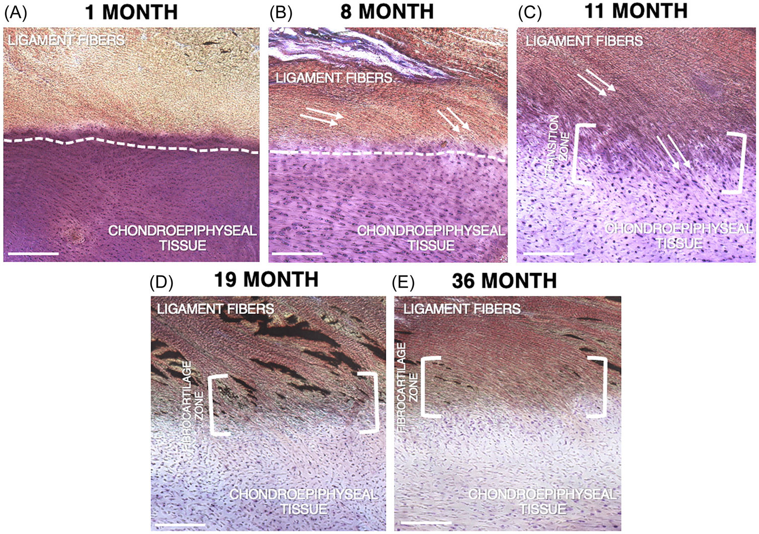
This study aimed to investigate the ultrastructural anatomy of the developing ACL tibial enthesis. We hypothesized that enthesis architecture would progressively mature and remodel, eventually resembling that of the adult by the early postnatal stage. Five fresh-frozen human pediatric cadaveric knees aged 1-36 months underwent anatomical dissection to harvest the ACL insertion and underlying tibial chondroepiphysis. The samples were prepared for scanning electron microscopy (SEM) to examine the ultrastructural anatomy of the enthesis and underwent histological staining for circular polarized light (CPL) and light microscopy imaging. SEM analysis of the 1- and 8-month-old samples revealed a shallow interdigitation between the dense fibrous (ligamentous) tissue and unmineralized chondrogenic tissues, with a minimal transition zone. By 11-month, a more complex transition zone was present. By age 19- and 36-month-old, a progressively more complex and defined fibrocartilage zone was observed. CPL analysis revealed distinct collagen fiber continuity, alignment, and organization changes over time. By 19 and 36 months, the samples exhibited complex fiber arrangements and a progression toward uniform fiber orientation. Similarly, histological analysis demonstrated progressive remodeling of the enthesis with increasing age. Our results suggest that the ACL enthesis of the developing knee begins to mimic that of an adult as early as 19 months of age, as a more complex transition between ligamentous and chondro-epiphyseal tissue can be appreciated. We hypothesize that the observed changes are likely due to mechanical loading of the enthesis with the onset of weightbearing. Future investigations of ACL reconstruction and repair will benefit from improved understanding of the chondro-epiphyseal/ACL regions.
本研究旨在调查发育中的前交叉韧带(ACL)胫骨附着点的超微结构解剖。我们假设附着点结构会逐渐成熟并重塑,最终在出生后早期类似于成年人的结构。对5个年龄在1至36个月的新鲜冷冻人类小儿尸体膝关节进行解剖,以获取ACL附着点及下方的胫骨软骨骨骺。制备样本用于扫描电子显微镜(SEM)检查附着点的超微结构解剖,并进行圆偏振光(CPL)组织学染色和光学显微镜成像。对1个月和8个月大样本的SEM分析显示,致密纤维(韧带)组织与未矿化软骨生成组织之间的指状交叉较浅,过渡区最小。到11个月时,出现了更复杂的过渡区。到19个月和36个月大时,观察到纤维软骨区逐渐变得更加复杂和明确。CPL分析显示,随着时间推移,胶原纤维的连续性、排列和组织发生了明显变化。到19个月和36个月时,样本呈现出复杂的纤维排列,并朝着纤维方向均匀化发展。同样,组织学分析表明,随着年龄增长,附着点逐渐重塑。我们的结果表明,发育中的膝关节的ACL附着点早在19个月大时就开始模仿成年人的附着点,因为可以观察到韧带组织和软骨骨骺组织之间有更复杂的过渡。我们推测观察到的变化可能是由于负重开始时附着点受到机械负荷所致。对ACL重建和修复的未来研究将受益于对软骨骨骺/ACL区域的更好理解。