Endodontic Department, Faculty of Dentistry, Future University in Egypt, New Cairo City, Egypt.
Ain Shams University, Cairo, Egypt.
Sci Rep. 2024 Oct 25;14(1):25378. doi: 10.1038/s41598-024-66033-w.
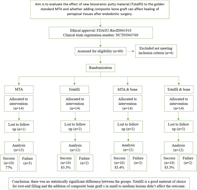
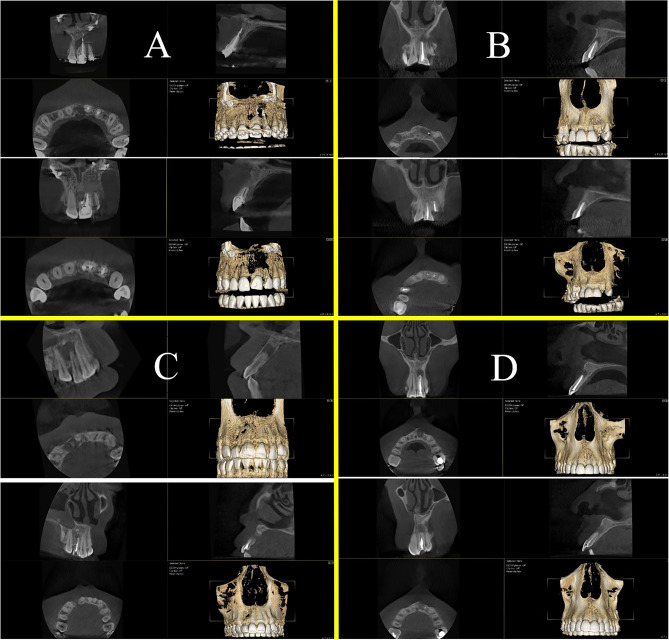
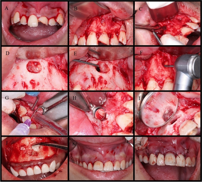
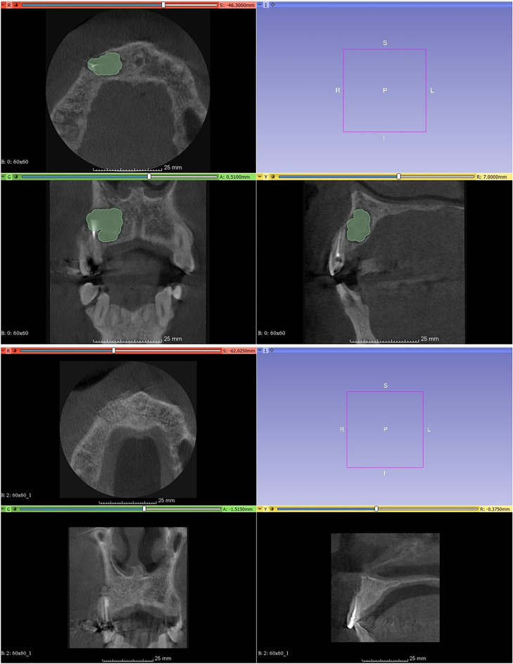
To evaluate the effect of combining different bioactive root-end filling materials with composite bone graft (xenogeneic mixed with autogenous bone fragments) on the healing process of periapical tissues after endodontic micro-surgery procedure. In this triple-blinded clinical trial, 56 patients were divided into 2 main groups (28 each) according to the root-end filling material and 2 subgroups according to the presence or absence of the composite bone graft material. Group I: MTA root-end filling (n = 28) in which there are Sub-group A: without bone graft (n = 14) and Sub-group B: with composite bone graft (n = 14). Group II: TotalFill root-end filling (n = 28) in which there are Sub-group A: without bone graft (n = 14) and Sub-group B: with composite bone graft (n = 14). Healthy patients whose ages range from 20 to 50 years with small-to-medium size radiolucency in CBCT related to single-rooted maxillary teeth were included in this study. Patients were assigned a number starting from 1 to 56 and were randomly allocated to four test groups (2 main groups and 2 sub-groups) following simple randomization procedure guidelines described by IBM SPSS V23 (IBM USA) statistical analysis software. This trial was triple-blind where the patient, the outcome assessors, and the main operator were blinded to the applied intervention. Every patient was evaluated clinically and by CBCT at two main observation periods: presurgical and 12-month post-operative. They were also examined and evaluated clinically and radiographically through periapical x-rays after one week, three, and six months. Statistical analysis was performed with IBM SPSS Statistics for Windows Version 23.0. Armonk, NY: IBM Corp. Of the 56 patients enrolled in the study, 49 patients were available for the final analysis. All groups showed no statistically significant differences with regard to healing or success rates at the 12-month follow-up mark. No adverse effects were encountered. Results showed that high success rates were achieved using MTA and TotalFill in the healing of periapical lesions after endodontic surgery. The addition of bone graft in small-to-medium size lesions did not affect the success rate of endodontic surgeries.
评估在根管显微手术中,不同生物活性根充材料与复合骨移植(异种混合自体骨碎片)联合使用对根尖组织愈合过程的影响。在这项三盲临床试验中,56 名患者根据根充材料分为 2 个主要组(每组 28 名),并根据是否存在复合骨移植材料进一步分为 2 个亚组。第 I 组:MTA 根充(n=28),其中亚组 A:无骨移植(n=14),亚组 B:复合骨移植(n=14)。第 II 组:TotalFill 根充(n=28),其中亚组 A:无骨移植(n=14),亚组 B:复合骨移植(n=14)。纳入研究的患者为年龄在 20 至 50 岁之间的健康患者,CBCT 显示上颌单根牙齿的小至中等大小的放射性不透明区与该研究相关。患者被分配从 1 到 56 的编号,并根据 IBM SPSS V23(美国 IBM)统计分析软件描述的简单随机化程序指南随机分配到四个测试组(2 个主要组和 2 个亚组)。该试验为三盲试验,患者、结果评估者和主要操作者均对所应用的干预措施不知情。每位患者均在两个主要观察期内进行临床和 CBCT 评估:术前和术后 12 个月。在术后一周、三个月和六个月,还通过根尖 X 线对患者进行了临床和影像学检查和评估。采用 IBM SPSS Statistics for Windows Version 23.0 进行统计分析。Armonk,NY:IBM Corp。在纳入研究的 56 名患者中,有 49 名患者可进行最终分析。所有组在 12 个月的随访标记时在愈合或成功率方面均无统计学差异。未出现不良反应。结果表明,在根管显微手术后,MTA 和 TotalFill 可实现较高的根尖病变愈合成功率。在小至中等大小病变中添加骨移植不会影响根管手术的成功率。