Brain Tumor Center, Erasmus MC Cancer Institute, University Medical Center Rotterdam, Rotterdam, The Netherlands.
Department of Radiotherapy, Erasmus MC Cancer Institute, University Medical Center Rotterdam, Rotterdam, The Netherlands.
Eur Radiol Exp. 2024 Oct 30;8(1):123. doi: 10.1186/s41747-024-00523-4.
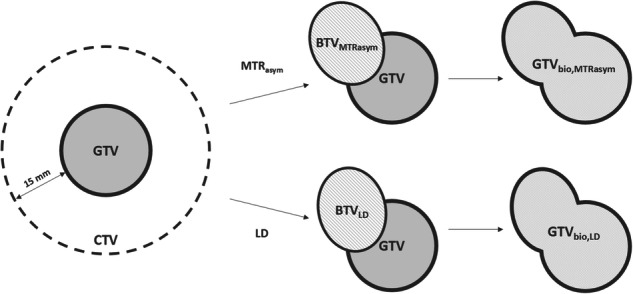
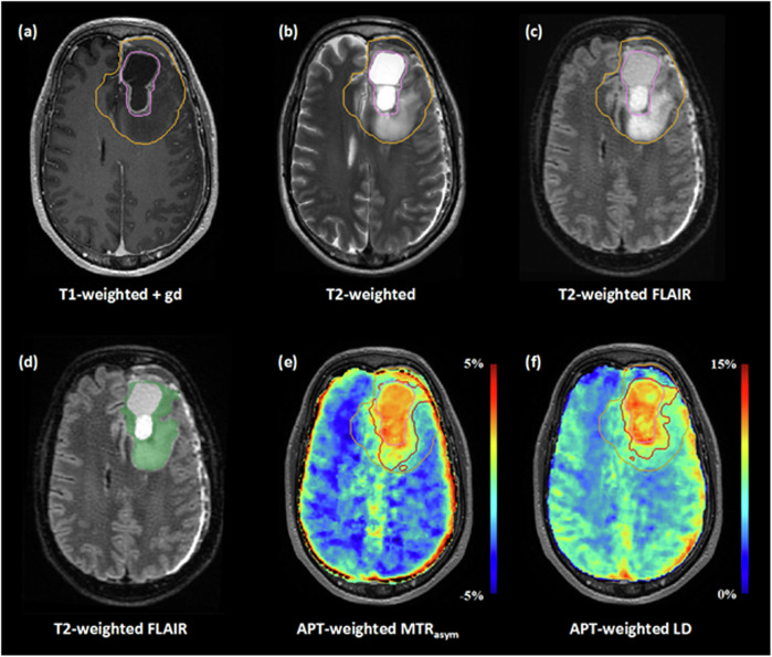
Extensive glioblastoma infiltration justifies a 15-mm margin around the gross tumor volume (GTV) to define the radiotherapy clinical target volume (CTV). Amide proton transfer (APT)-weighted imaging could enable visualization of tumor infiltration, allowing more accurate GTV delineation. We quantified the impact of integrating APT-weighted imaging into GTV delineation of glioblastoma and compared two APT-weighted quantification methods-magnetization transfer ratio asymmetry (MTR) and Lorentzian difference (LD) analysis-for target delineation.
Nine glioblastoma patients underwent an extended imaging protocol prior to radiotherapy, yielding APT-weighted MTR and LD maps. From both maps, biological tumor volumes were generated (BTV and BTV) and added to the conventional GTV to generate biological GTVs (GTV and GTV). Wilcoxon signed-rank tests were performed for comparisons.
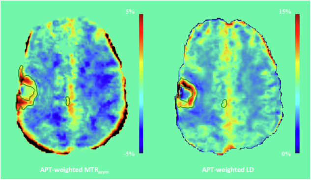
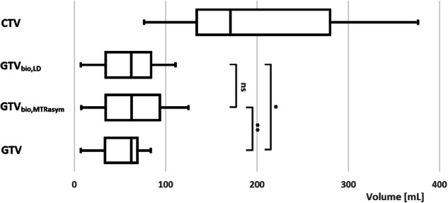
The GTV and GTV were significantly larger than the conventional GTV (p ≤ 0.022), with a median volume increase of 9.3% and 2.1%, respectively. The GTV and GTV were significantly smaller than the CTV (p = 0.004), with a median volume reduction of 72.1% and 70.9%, respectively. There was no significant volume difference between the BTV and BTV (p = 0.074). In three patients, BTV delineation was affected by elevated signals at the brain periphery due to residual motion artifacts; this elevation was absent on the APT-weighted LD maps.
Larger biological GTVs compared to the conventional GTV highlight the potential of APT-weighted imaging for radiotherapy target delineation of glioblastoma. APT-weighted LD mapping may be advantageous for target delineation as it may be more robust against motion artifacts.
The introduction of APT-weighted imaging may, ultimately, enhance visualization of tumor infiltration and eliminate the need for the substantial 15-mm safety margin for target delineation of glioblastoma. This could reduce the risk of radiation toxicity while still effectively irradiating the tumor.
NCT05970757 (ClinicalTrials.gov).
Integration of APT-weighted imaging into target delineation for radiotherapy is feasible. The integration of APT-weighted imaging yields larger GTVs in glioblastoma. APT-weighted LD mapping may be more robust against motion artifacts than APT-weighted MTR.
广泛的胶质母细胞瘤浸润使得在大体肿瘤体积(GTV)周围需要 15mm 的边界来定义放射治疗临床靶区(CTV)。酰胺质子转移(APT)加权成像可以实现肿瘤浸润的可视化,从而更准确地描绘 GTV。我们量化了将 APT 加权成像整合到胶质母细胞瘤 GTV 描绘中的影响,并比较了两种 APT 加权定量方法——磁化转移率不对称(MTR)和洛伦兹差值(LD)分析——在靶区描绘中的作用。
9 名胶质母细胞瘤患者在放疗前接受了扩展成像方案,获得了 APT 加权 MTR 和 LD 图。从这两个图中,生成了生物肿瘤体积(BTV 和 BTV)并添加到常规 GTV 中,生成了生物 GTV(GTV 和 GTV)。使用 Wilcoxon 符号秩检验进行比较。
GTV 和 GTV 明显大于常规 GTV(p≤0.022),体积分别增加了 9.3%和 2.1%。GTV 和 GTV 明显小于 CTV(p=0.004),体积分别减少了 72.1%和 70.9%。BTV 和 BTV 之间的体积没有显著差异(p=0.074)。在 3 名患者中,由于残留运动伪影,脑周边的升高信号影响了 BTV 描绘;而在 APT 加权 LD 图上则不存在这种升高。
与常规 GTV 相比,更大的生物 GTV 突出了 APT 加权成像在胶质母细胞瘤放疗靶区描绘中的潜力。APT 加权 LD 映射可能在靶区描绘方面具有优势,因为它可能对运动伪影更稳健。
APT 加权成像的引入最终可能会增强肿瘤浸润的可视化,并消除胶质母细胞瘤靶区描绘中对 15mm 安全边界的大量需求。这可以降低放射性毒性的风险,同时仍然有效地照射肿瘤。
NCT05970757(ClinicalTrials.gov)。
将 APT 加权成像整合到放疗靶区描绘中是可行的。APT 加权成像在胶质母细胞瘤中产生更大的 GTV。APT 加权 LD 映射可能比 APT 加权 MTR 更能抵抗运动伪影。