Prykhozhij Sergey V, Ban Kevin, Brown Zane L, Kobar Kim, Wajnberg Gabriel, Fuller Charlotte, Chacko Simi, Lacroix Jacynthe, Crapoulet Nicolas, Midgen Craig, Shlien Adam, Malkin David, Berman Jason N
Children's Hospital of Eastern Ontario (CHEO) Research Institute and University of Ottawa, Ottawa, ON, Canada.
Dalhousie University Medical School, Halifax, NS, Canada.
Data Brief. 2024 Oct 16;57:111041. doi: 10.1016/j.dib.2024.111041. eCollection 2024 Dec.
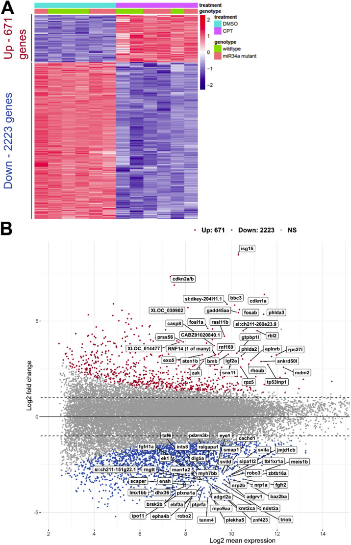
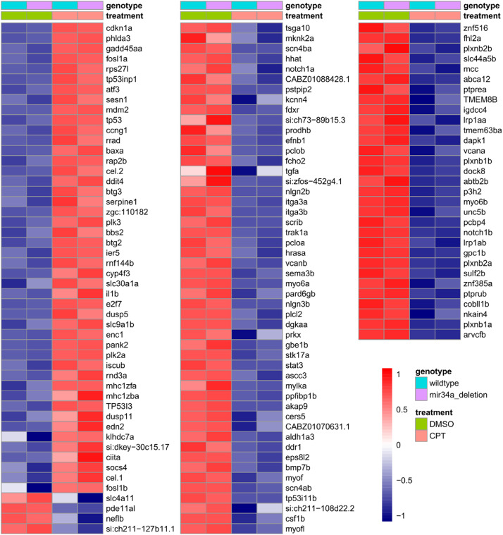
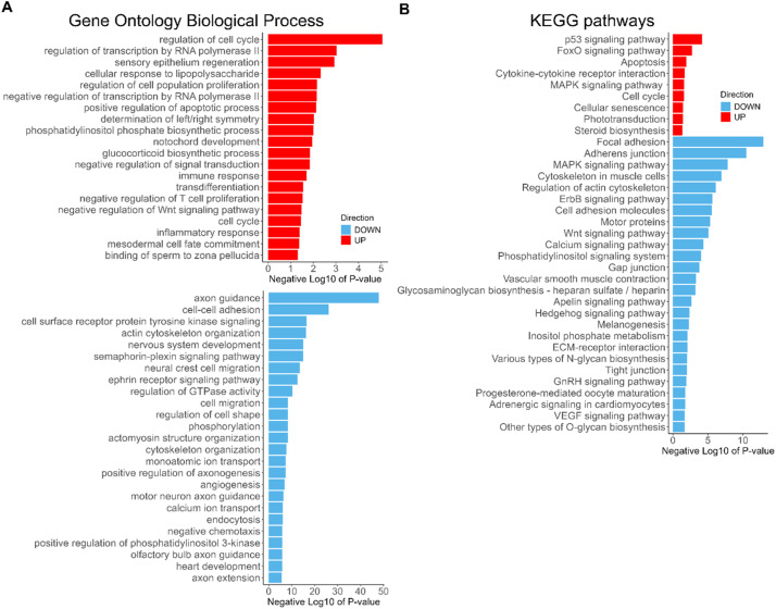
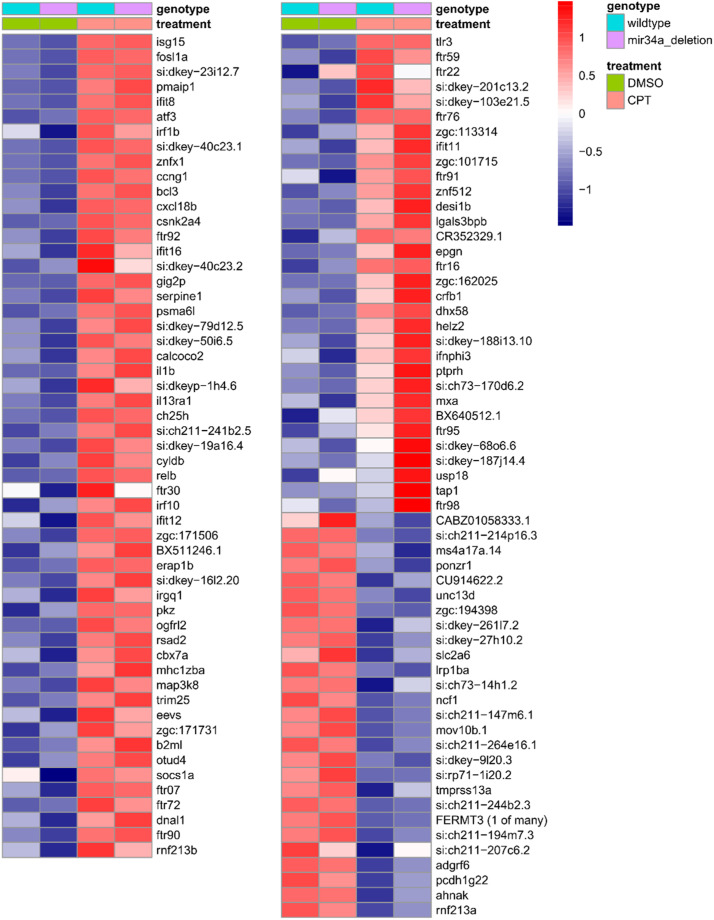
Zebrafish () are a good model for cancer research including studies on chemotherapy treatments. We treated wild-type and deletion mutant zebrafish embryos at 24 h post-fertilization with 1 µM of the topoisomerase I inhibitor, camptothecin (CPT), for 4 h to catalogue gene expression changes induced by this DNA damage treatment and to understand if these changes are influenced by loss of miR-34a. The 4 sample groups of 3 independent biological samples consisting of 30 embryos each were analyzed by RNA-sequencing using the recently updated zebrafish transcriptome annotation based on GRCz11, which enabled a more complete and sensitive read mapping and gene assignment than standard annotations. Using this gene expression estimates dataset as the primary resource, we performed a differentially expressed gene (DEG) analysis based on treatment as loss of miR-34a had minimal effects on CPT-induced expression changes. The DEGs were analyzed for Gene Ontology and KEGG pathway terms. Enriched terms and pathways among up-regulated genes were mostly related to stress, cell death, cell cycle regulation, transcriptional regulation, cell signalling, developmental processes and synthesis of retinol and steroid hormones. By contrast, down-regulated genes were most strongly associated with genes involved in key developmental processes, adhesion molecules, as well as some transport and metabolic pathways, together suggesting a "developmental shutdown". We also identified interferon-regulated genes and p53 target genes activated or inhibited by DNA damage due to topoisomerase I inhibition, suggesting that they are important components of the response to this type of DNA damage in zebrafish embryos.
斑马鱼是癌症研究的良好模型,包括化疗治疗研究。我们在受精后24小时用1μM的拓扑异构酶I抑制剂喜树碱(CPT)处理野生型和miR-34a缺失突变体斑马鱼胚胎4小时,以梳理这种DNA损伤处理诱导的基因表达变化,并了解这些变化是否受miR-34a缺失的影响。由30个胚胎组成的4个样本组,每组有3个独立的生物学样本,使用基于GRCz11的最新更新的斑马鱼转录组注释通过RNA测序进行分析,与标准注释相比,这使得读取映射和基因分配更加完整和敏感。以这个基因表达估计数据集作为主要资源,我们基于处理进行了差异表达基因(DEG)分析,因为miR-34a的缺失对CPT诱导的表达变化影响最小。对差异表达基因进行了基因本体论和KEGG通路术语分析。上调基因中富集的术语和通路大多与应激、细胞死亡、细胞周期调控、转录调控、细胞信号传导、发育过程以及视黄醇和类固醇激素的合成有关。相比之下,下调基因与参与关键发育过程的基因、粘附分子以及一些运输和代谢通路密切相关,共同表明出现了“发育停滞”。我们还鉴定了因拓扑异构酶I抑制导致DNA损伤而被激活或抑制的干扰素调节基因和p53靶基因,表明它们是斑马鱼胚胎对这种类型DNA损伤反应的重要组成部分。