文献检索文档翻译深度研究
Suppr Zotero 插件Zotero 插件
邀请有礼套餐&价格历史记录

新学期,新优惠

限时优惠:9月1日-9月22日

30天高级会员仅需29元

1天体验卡首发特惠仅需5.99元

了解详情
不再提醒
插件&应用
Suppr Zotero 插件Zotero 插件浏览器插件Mac 客户端Windows 客户端微信小程序
高级版
套餐订阅购买积分包
AI 工具
文献检索文档翻译深度研究
关于我们
关于 Suppr公司介绍联系我们用户协议隐私条款
关注我们

Suppr 超能文献

核心技术专利:CN118964589B侵权必究
粤ICP备2023148730 号-1Suppr @ 2025

产前地塞米松暴露通过 Mkp-1 基因座上的组蛋白修饰减少小鼠成骨前体细胞的增殖。

Prenatal dexamethasone exposure reduces osteoprogenitor proliferation in mice via histone modifications at the Mkp-1 gene locus.

机构信息

Division of Orthopaedics and Traumatology, Department of Orthopaedics, Nanfang Hospital, Southern Medical University, No.1838 North of Guangzhou Avenue, Guangzhou, 510515, Guangdong Province, China.

Guangdong Provincial Key Laboratory of Bone and Cartilage Regenerative Medicine, Nanfang Hospital, Southern Medical University, Guangzhou, 510515, China.

出版信息

Commun Biol. 2024 Nov 28;7(1):1589. doi: 10.1038/s42003-024-07288-x.


DOI:10.1038/s42003-024-07288-x
PMID:39609620
原文链接:https://pmc.ncbi.nlm.nih.gov/articles/PMC11604782/
Abstract

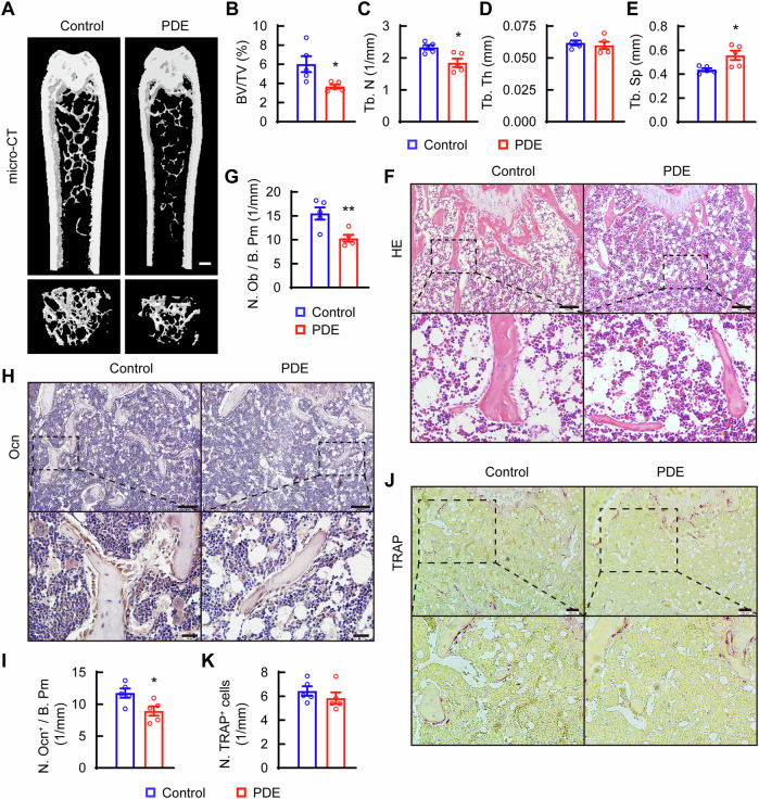
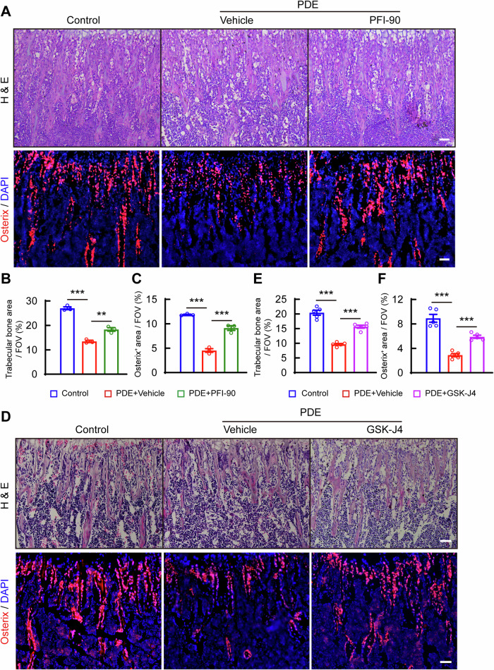
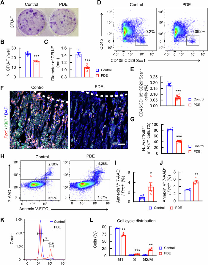
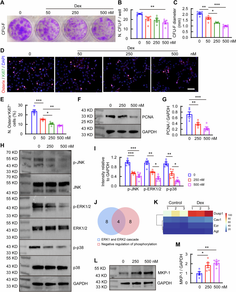
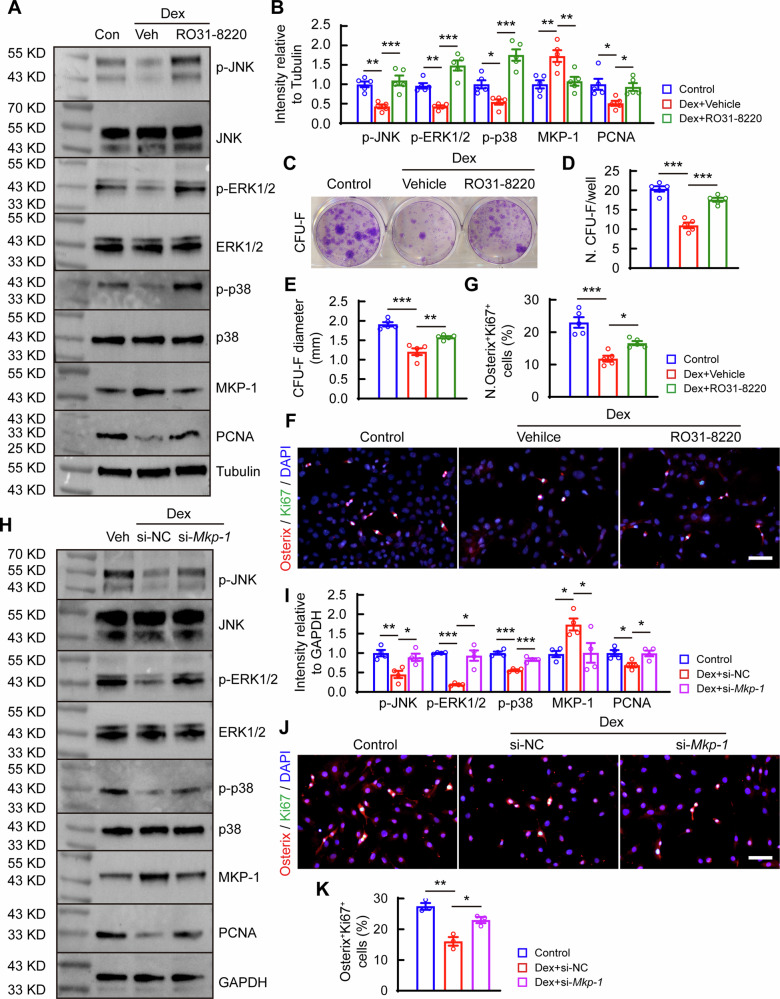
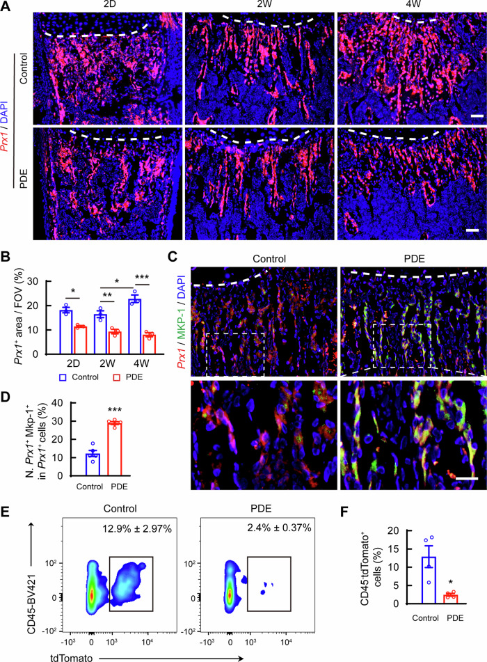
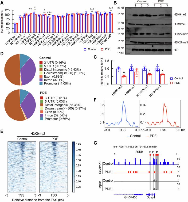
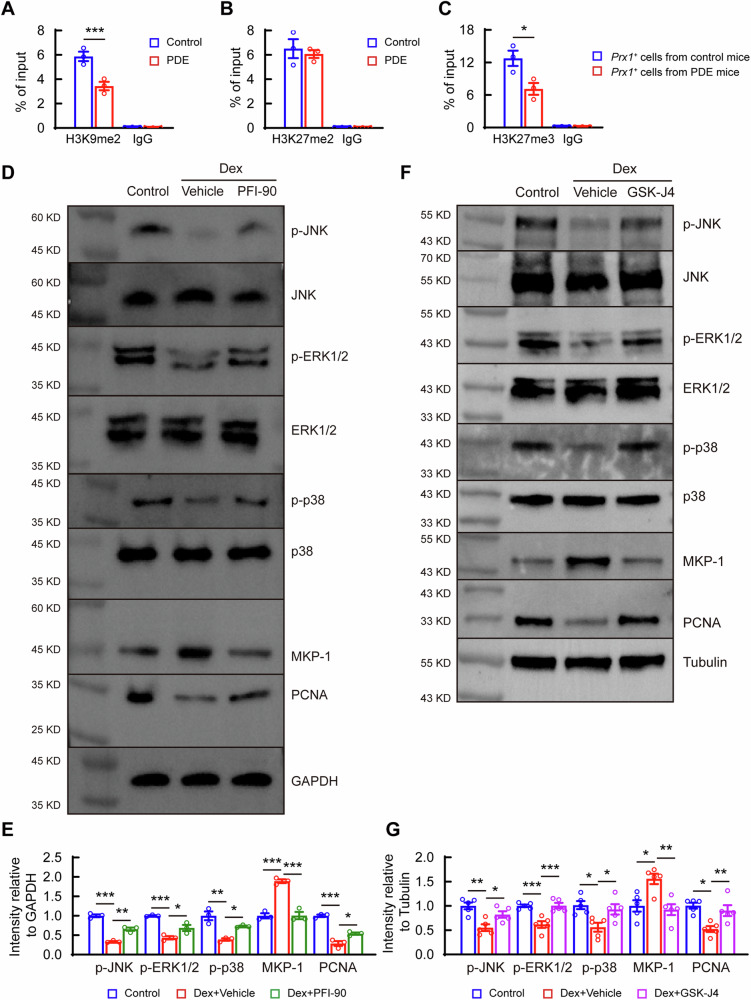
Prenatal dexamethasone exposure (PDE) has long-term consequences in bone development, though the underlying mechanisms remain unclear. Our results show that PDE offspring exhibit reduced bone mass, fewer osteoblasts and diminished osteoprogenitors proliferation. Further analyses show that PDE increases MKP-1 expression, while decreasing H3 lysine 9 dimethylation (H3K9me2) and H3 lysine 27 trimethylation (H3K27me3) at the Mkp-1 gene locus. Mechanistically, dexamethasone suppresses osteoprogenitors proliferation by upregulating MKP-1 expression, notably through the inhibition of H3K9me2 and H3K27me3 modifications, which promote demethylation and transcriptional activation of the Mkp-1 gene. Importantly, restoring histone methylation balance with PFI-90 or GSK-J4 treatment blocks the inhibitory effects of PDE on MAPK signaling in osteoprogenitors, and mitigates the detrimental impact of PDE on osteoprogenitor proliferation and bone development in the offspring. This study provides new insights into the epigenetic mechanism by which PDE disrupts long-term programming of fetal osteoprogenitor proliferation, ultimately impairing long bone growth in offspring.

摘要

产前地塞米松暴露(PDE)对骨骼发育有长期影响,但潜在机制尚不清楚。我们的结果表明,PDE 后代表现出骨量减少、成骨细胞减少和骨祖细胞增殖减少。进一步的分析表明,PDE 增加了 MKP-1 的表达,同时降低了 Mkp-1 基因座上的 H3 赖氨酸 9 二甲基化(H3K9me2)和 H3 赖氨酸 27 三甲基化(H3K27me3)。从机制上讲,地塞米松通过上调 MKP-1 的表达来抑制骨祖细胞的增殖,特别是通过抑制 H3K9me2 和 H3K27me3 修饰,促进 Mkp-1 基因的去甲基化和转录激活。重要的是,用 PFI-90 或 GSK-J4 治疗恢复组蛋白甲基化平衡可阻断 PDE 对骨祖细胞中 MAPK 信号的抑制作用,并减轻 PDE 对后代骨祖细胞增殖和骨发育的有害影响。这项研究为 PDE 破坏胎儿骨祖细胞增殖的长期编程的表观遗传机制提供了新的见解,最终损害了后代长骨的生长。

https://cdn.ncbi.nlm.nih.gov/pmc/blobs/d8b5/11604782/044292ee5281/42003_2024_7288_Fig8_HTML.jpg
https://cdn.ncbi.nlm.nih.gov/pmc/blobs/d8b5/11604782/ddbeb0cd9c88/42003_2024_7288_Fig1_HTML.jpg
https://cdn.ncbi.nlm.nih.gov/pmc/blobs/d8b5/11604782/d5348217df46/42003_2024_7288_Fig2_HTML.jpg
https://cdn.ncbi.nlm.nih.gov/pmc/blobs/d8b5/11604782/1afd796ba000/42003_2024_7288_Fig3_HTML.jpg
https://cdn.ncbi.nlm.nih.gov/pmc/blobs/d8b5/11604782/a4a255f63063/42003_2024_7288_Fig4_HTML.jpg
https://cdn.ncbi.nlm.nih.gov/pmc/blobs/d8b5/11604782/6d77af38f631/42003_2024_7288_Fig5_HTML.jpg
https://cdn.ncbi.nlm.nih.gov/pmc/blobs/d8b5/11604782/78b3ad8a73f2/42003_2024_7288_Fig6_HTML.jpg
https://cdn.ncbi.nlm.nih.gov/pmc/blobs/d8b5/11604782/99efedc22b78/42003_2024_7288_Fig7_HTML.jpg
https://cdn.ncbi.nlm.nih.gov/pmc/blobs/d8b5/11604782/044292ee5281/42003_2024_7288_Fig8_HTML.jpg

相似文献

[1]
Prenatal dexamethasone exposure reduces osteoprogenitor proliferation in mice via histone modifications at the Mkp-1 gene locus.

Commun Biol. 2024-11-28

[2]
Mitogen-activated protein kinase phosphatase 1/dual specificity phosphatase 1 mediates glucocorticoid inhibition of osteoblast proliferation.

Mol Endocrinol. 2007-12

[3]
Prenatal dexamethasone exposure impairs rat blood-testis barrier function and sperm quality in adult offspring via GR/KDM1B/FSTL3/TGFβ signaling.

Acta Pharmacol Sin. 2024-6

[4]
Increased H3K27ac level of ACE mediates the intergenerational effect of low peak bone mass induced by prenatal dexamethasone exposure in male offspring rats.

Cell Death Dis. 2018-5-29

[5]
The Role of MKP-1 in the Anti-Proliferative Effects of Glucocorticoids in Primary Rat Pre-Osteoblasts.

PLoS One. 2015-8-11

[6]
11β-Hydroxysteroid dehydrogenase 2: A key mediator of high susceptibility to osteoporosis in offspring after prenatal dexamethasone exposure.

Pharmacol Res. 2022-1

[7]
Decreased H3K9ac level of AT2R mediates the developmental origin of glomerulosclerosis induced by prenatal dexamethasone exposure in male offspring rats.

Toxicology. 2019-1-1

[8]
Mitogen-activated protein kinase phosphatase-1 promotes neovascularization and angiogenic gene expression.

Arterioscler Thromb Vasc Biol. 2014-2-27

[9]
Low H3K27 acetylation of SF1 in PBMC: a biomarker for prenatal dexamethasone exposure-caused adrenal insufficiency of steroid synthesis in male offspring.

Cell Biol Toxicol. 2023-10

[10]
Course-, dose-, and stage-dependent toxic effects of prenatal dexamethasone exposure on long bone development in fetal mice.

Toxicol Appl Pharmacol. 2018-5-9

本文引用的文献

[1]
Depletion of SAM leading to loss of heterochromatin drives muscle stem cell ageing.

Nat Metab. 2024-1

[2]
SRplot: A free online platform for data visualization and graphing.

PLoS One. 2023

[3]
Oxylipin-PPARγ-initiated adipocyte senescence propagates secondary senescence in the bone marrow.

Cell Metab. 2023-4-4

[4]
Prrx1 marks stem cells for bone, white adipose tissue and dermis in adult mice.

Nat Genet. 2022-12

[5]
Different corticosteroids and regimens for accelerating fetal lung maturation for babies at risk of preterm birth.

Cochrane Database Syst Rev. 2022-8-9

[6]
Recent Advances in Single-Cell View of Mesenchymal Stem Cell in Osteogenesis.

Front Cell Dev Biol. 2022-1-5

[7]
The psychological distress of parents is associated with reduced linear growth of children: Evidence from a nationwide population survey.

PLoS One. 2021

[8]
Cell-Type-Specific Impact of Glucocorticoid Receptor Activation on the Developing Brain: A Cerebral Organoid Study.

Am J Psychiatry. 2022-5

[9]
Interplay between chromatin marks in development and disease.

Nat Rev Genet. 2022-3

[10]
Non-autonomous regulation of germline stem cell proliferation by somatic MPK-1/MAPK activity in C. elegans.

Cell Rep. 2021-5-25

文献AI研究员

20分钟写一篇综述,助力文献阅读效率提升50倍

立即体验

用中文搜PubMed

大模型驱动的PubMed中文搜索引擎

马上搜索

推荐工具

医学文档翻译智能文献检索