McIlvried Lisa A, Del Rosario John Smith, Pullen Melanie Y, Wangzhou Andi, Sheahan Tayler D, Shepherd Andrew J, Slivicki Richard A, Lemen John A, Price Theodore J, Copits Bryan A, Gereau Robert W
Washington University Pain Center and Department of Anesthesiology, Washington University School of Medicine, St. Louis, MO, USA.
Department of Neuroscience and Center for Advanced Pain Studies, The University of Texas at Dallas, Dallas, TX, USA.
J Gen Physiol. 2025 Jan 6;157(1). doi: 10.1085/jgp.202313488. Epub 2024 Dec 17.
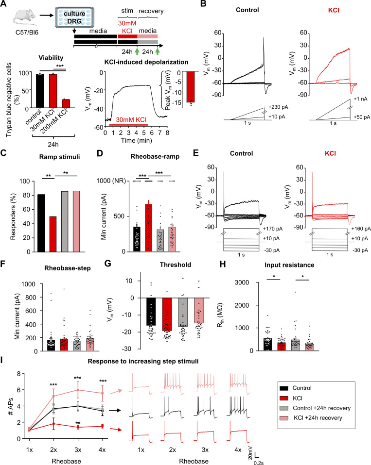
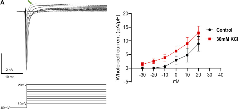
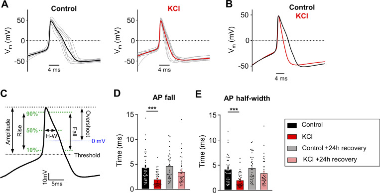
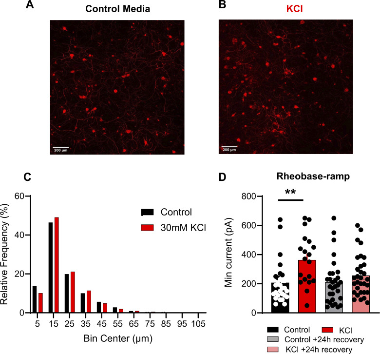
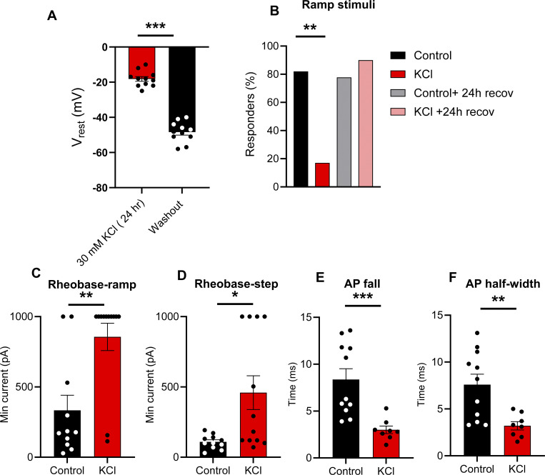
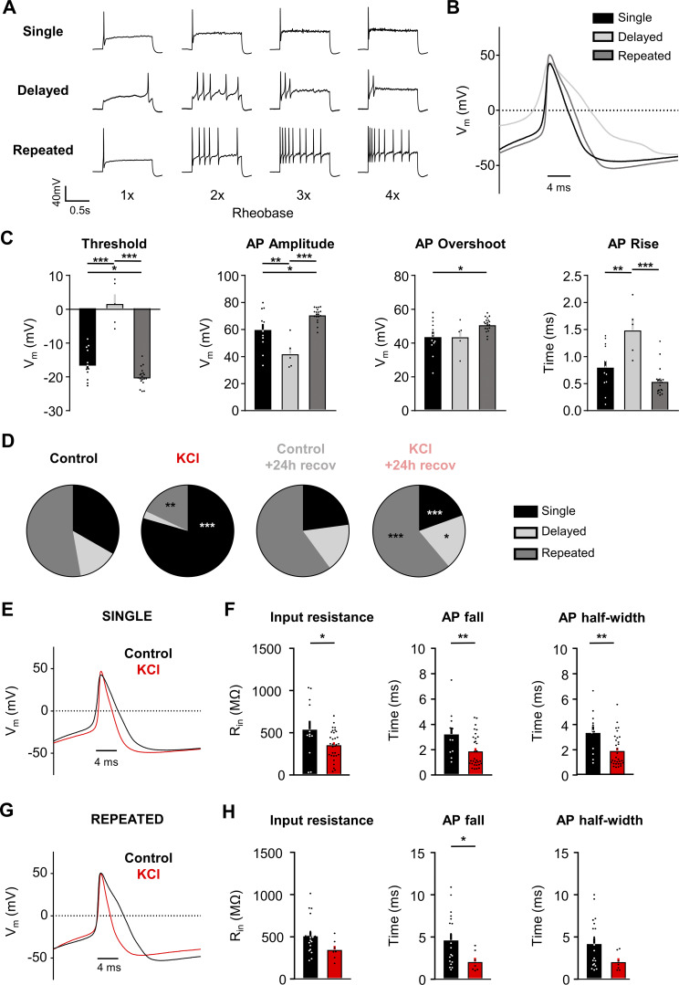
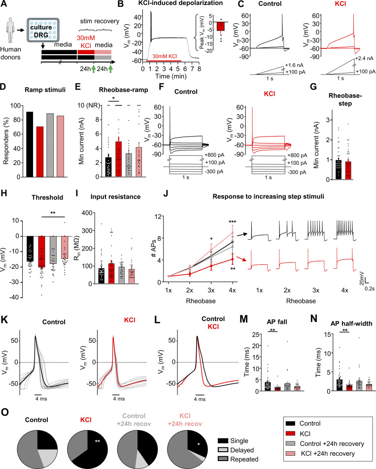
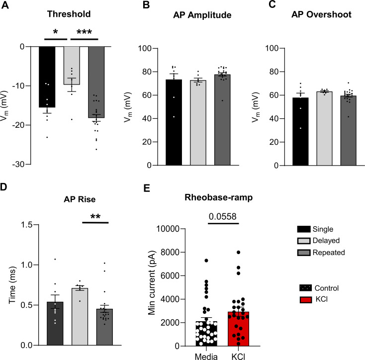
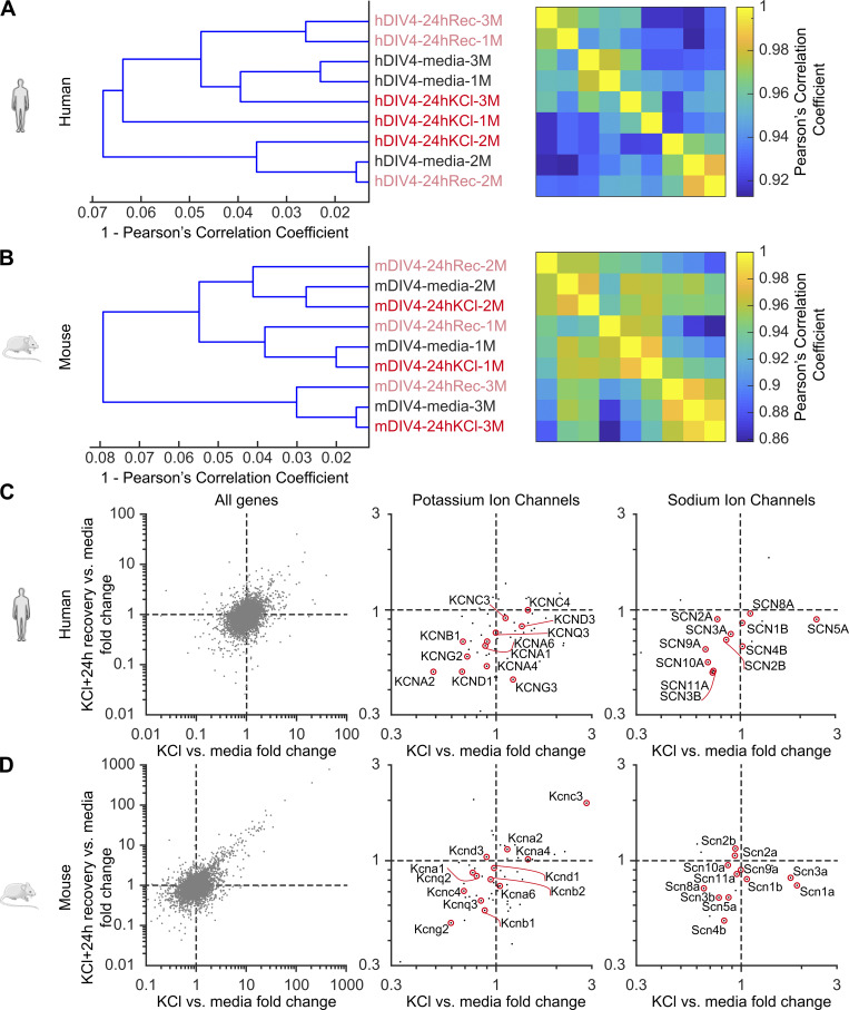
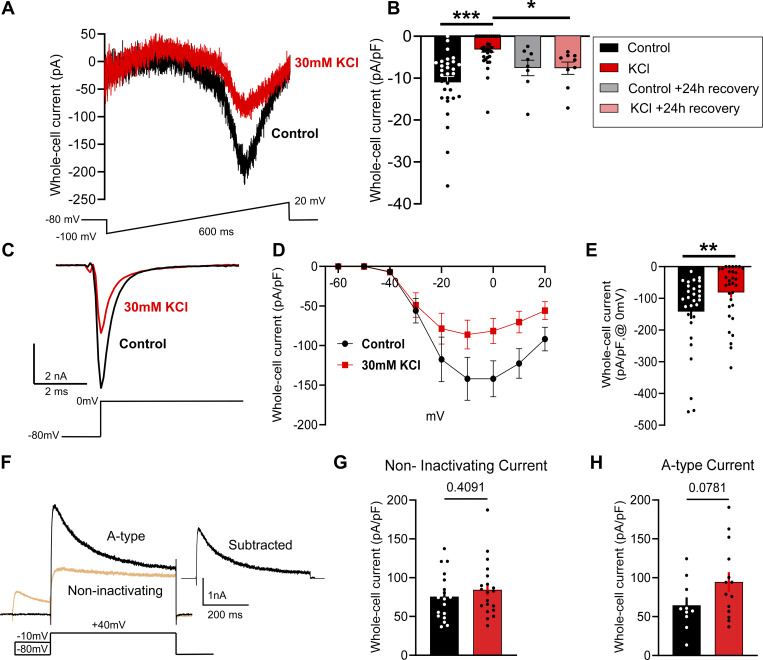
In response to changes in activity induced by environmental cues, neurons in the central nervous system undergo homeostatic plasticity to sustain overall network function during abrupt changes in synaptic strengths. Homeostatic plasticity involves changes in synaptic scaling and regulation of intrinsic excitability. Increases in spontaneous firing and excitability of sensory neurons are evident in some forms of chronic pain in animal models and human patients. However, whether mechanisms of homeostatic plasticity are engaged in sensory neurons of the peripheral nervous system (PNS) is unknown. Here, we show that sustained depolarization (induced by 24-h incubation in 30 mM KCl) induces compensatory changes that decrease the excitability of mouse and human sensory neurons without directly opposing membrane depolarization. Voltage-clamp recordings show that sustained depolarization produces no significant alteration in voltage-gated potassium currents, but a robust reduction in voltage-gated sodium currents, likely contributing to the overall decrease in neuronal excitability. The compensatory decrease in neuronal excitability and reduction in voltage-gated sodium currents reversed completely following a 24-h recovery period in a normal medium. Similar adaptive changes were not observed in response to 24 h of sustained action potential firing induced by optogenetic stimulation at 1 Hz, indicating the need for prolonged depolarization to drive engagement of this adaptive mechanism in sensory neurons. Our findings show that mouse and human sensory neurons are capable of engaging adaptive mechanisms to regulate intrinsic excitability in response to sustained depolarization in a manner similar to that described in neurons in the central nervous system.
为响应环境线索诱导的活动变化,中枢神经系统中的神经元会经历稳态可塑性,以在突触强度突然变化期间维持整体网络功能。稳态可塑性涉及突触缩放的变化和内在兴奋性的调节。在动物模型和人类患者的某些慢性疼痛形式中,感觉神经元的自发放电和兴奋性增加是明显的。然而,稳态可塑性机制是否在外周神经系统(PNS)的感觉神经元中起作用尚不清楚。在这里,我们表明持续去极化(由在30 mM KCl中孵育24小时诱导)会诱导补偿性变化,从而降低小鼠和人类感觉神经元的兴奋性,而不会直接对抗膜去极化。电压钳记录显示,持续去极化不会使电压门控钾电流产生显著改变,但会使电压门控钠电流显著降低,这可能导致神经元兴奋性的整体下降。在正常培养基中恢复24小时后,神经元兴奋性的补偿性降低和电压门控钠电流的减少完全逆转。在1 Hz光遗传学刺激诱导的24小时持续动作电位发放后,未观察到类似的适应性变化,这表明需要长时间去极化来驱动这种适应性机制在感觉神经元中的参与。我们的研究结果表明,小鼠和人类感觉神经元能够以类似于中枢神经系统中神经元所描述的方式,参与适应性机制来调节内在兴奋性,以响应持续去极化。