Santanda Takushi, Nakamura Yuichi, Ito Joji
Department of Critical Care Medicine, Itabashi Chuo Medical Center, Itabashi, JPN.
Department of Emergency and Critical Care Medicine, Tokyo Bay Urayasu Ichikawa Medical Center, Urayasu, JPN.
Cureus. 2024 Nov 18;16(11):e73942. doi: 10.7759/cureus.73942. eCollection 2024 Nov.
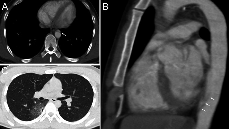
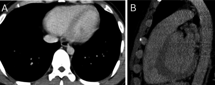
Contrast-enhanced CT is a primary tool in emergency departments for diagnosing acute aortic dissection, demonstrating high sensitivity and specificity. However, artifacts such as streak artifacts can mimic aortic dissection, leading to misdiagnosis. Here, we report a case involving a 21-year-old male who sustained traumatic injuries after a motor vehicle accident. Initial contrast-enhanced CT indicated a possible localized dissection in the descending aorta. Conservative treatment was initiated under the presumption of aortic dissection. Upon re-evaluation with ECG-gated CT, the previously identified "dissection" artifact had disappeared, revealing no actual aortic injury. This case illustrates how heartbeat-induced streak artifacts, while commonly seen in the aortic root, can also manifest in the descending aorta. Our findings underscore the importance of considering artifacts in atypical cases of aortic dissection, particularly when findings are localized to areas of the aorta in close proximity to the heart. For trauma patients, while dynamic contrast-enhanced CT remains standard, ECG-gated CT should be selectively applied where motion artifacts are suspected. This case highlights the role of advanced imaging options in distinguishing between true aortic pathology and artifacts, aiding in appropriate clinical decision-making.
增强CT是急诊科诊断急性主动脉夹层的主要工具,具有较高的敏感性和特异性。然而,诸如条纹伪影等伪影可能会模拟主动脉夹层,导致误诊。在此,我们报告一例涉及一名21岁男性的病例,该男性在机动车事故后遭受创伤。初始增强CT显示降主动脉可能存在局限性夹层。在假定为主动脉夹层的情况下开始保守治疗。经心电图门控CT重新评估后,先前发现的“夹层”伪影消失,未发现实际的主动脉损伤。该病例说明了心跳引起的条纹伪影虽然常见于主动脉根部,但也可出现在降主动脉。我们的研究结果强调了在非典型主动脉夹层病例中考虑伪影的重要性,特别是当发现局限于主动脉靠近心脏的区域时。对于创伤患者,虽然动态增强CT仍然是标准检查,但在怀疑有运动伪影的情况下应选择性地应用心电图门控CT。该病例突出了先进成像方法在区分真正的主动脉病变和伪影方面的作用,有助于做出适当的临床决策。