Später Thomas, Del Rio Patricia, Shelest Oksana, Wechsler Jacob T, Kaneda Giselle, Chavez Melissa, Sheyn Julia, Yu Victoria, Metzger Wolfgang, Huang Dave, Metzger Melodie, Tawackoli Wafa, Sheyn Dmitriy
Orthopaedic Stem Cell Research Laboratory, Cedars-Sinai Medical Center, Los Angeles, CA, United States.
Board of Governors Regenerative Medicine Institute, Cedars-Sinai Medical Center, Los Angeles, CA, United States.
Front Bioeng Biotechnol. 2024 Dec 6;12:1407729. doi: 10.3389/fbioe.2024.1407729. eCollection 2024.
Tendon injuries represent an ongoing challenge in clinical practice due to poor regenerative capacity, structure, and biomechanical function recovery of ruptured tendons. This study is focused on the assessment of a novel strategy to repair ruptured Achilles tendons in a Nude rat model using stem cell-seeded biomaterial.
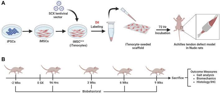
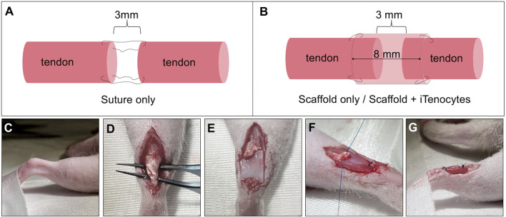
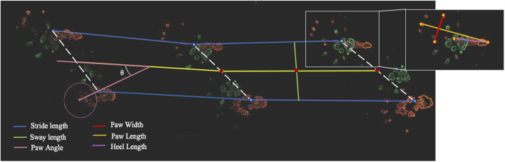
Specifically, we have used induced pluripotent stem cell (iPSC)-derived mesenchymal stem cells (iMSCs) overexpressing the early tendon marker Scleraxis (SCX, iMSC, iTenocytes) in combination with an elastic collagen scaffold. Achilles tendon defects in Nude rat models were created by isolating the tendon and excising 3 mm of the midsection. The Achilles tendon defects were then repaired with iTenocyte-seeded scaffolds, unseeded scaffolds, or suture only and compared to native Nude rat tendon tissue using gait analyses, biomechanical testing, histology, and immunohistochemistry.
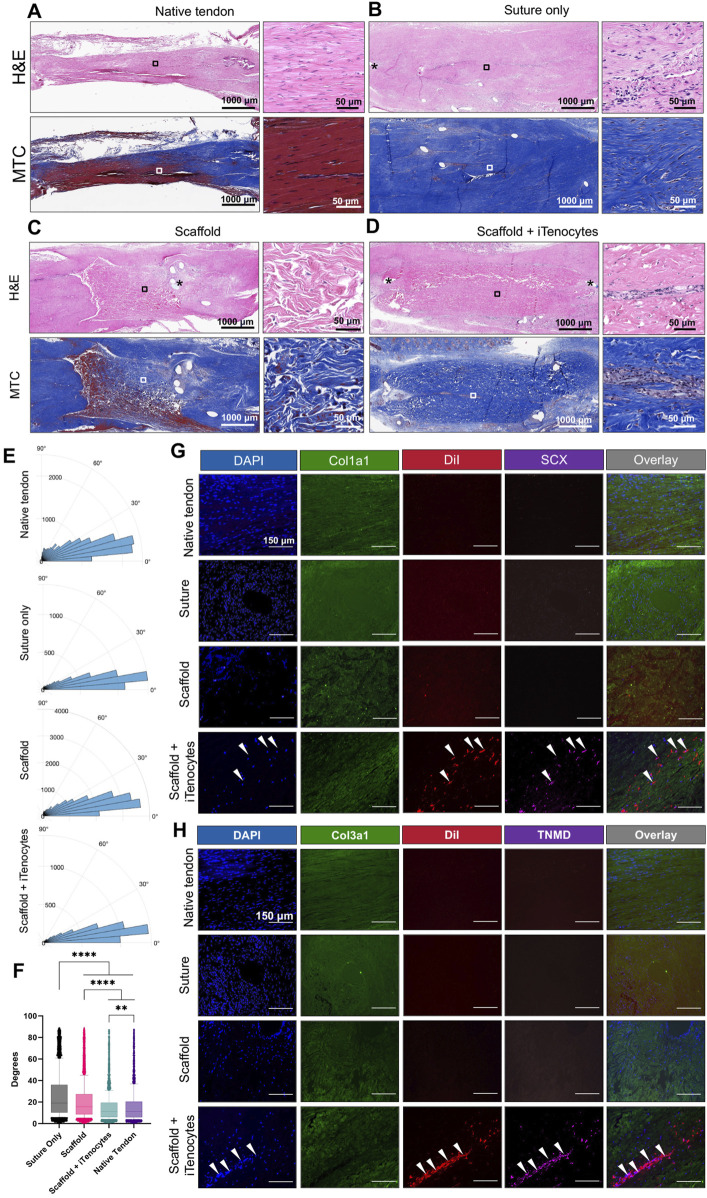
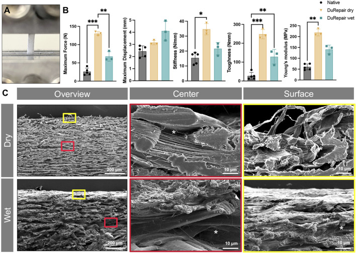
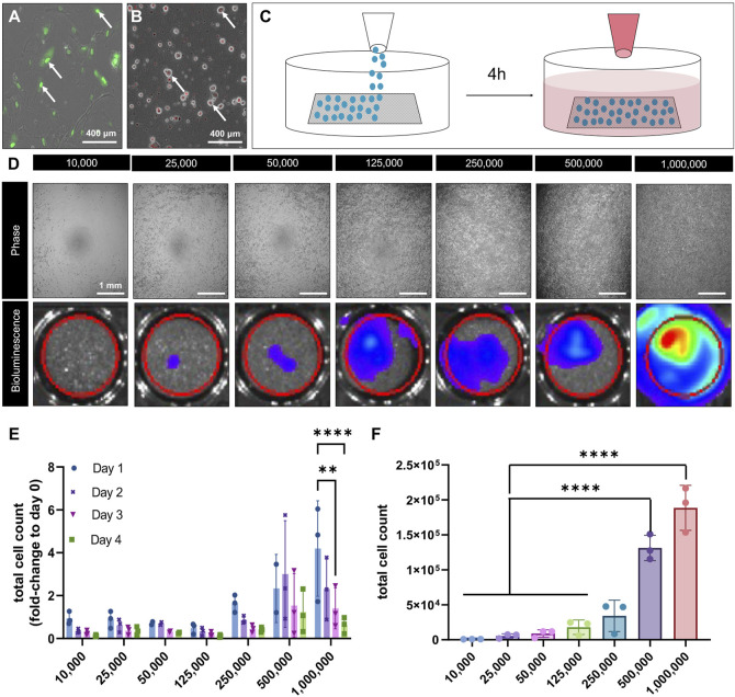
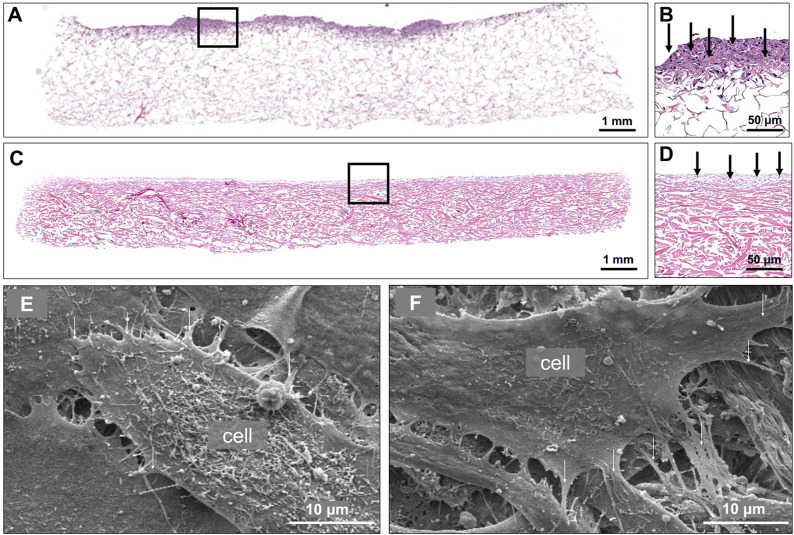
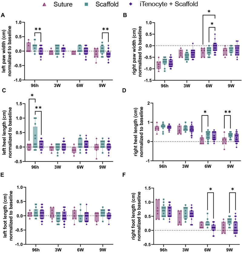
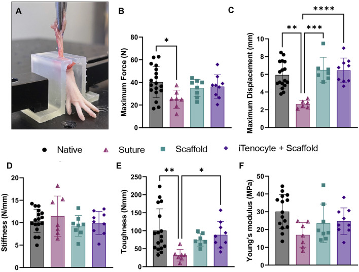
The results show faster functional recovery of gait in iTenocyte-seeded scaffold group comparing to scaffold only and suture only groups. Both iTenocyte-seeded scaffold and scaffold only treatment groups had improved biomechanical properties when compared to suture only treatment group, however no statistically significant difference was found in comparing the cell seeding scaffold an scaffold only group in terms of biomechanical properties. Immunohistochemistry staining further demonstrated that iTenocytes successfully populated the collagen scaffolds and survived 9 weeks after implantation . Additionally, the repaired tissue of iTenocyte-treated injuries exhibited a more organized structure when compared to tendon defects that were repaired only with suturing or unseeded scaffolds.
We suggest that iTenocyte-seeded DuRepair™ collagen scaffold can be used as potential treatment to regenerate the tendon tissue biomechanically and functionally.
由于断裂肌腱的再生能力差、结构和生物力学功能恢复不佳,肌腱损伤在临床实践中仍然是一个持续存在的挑战。本研究的重点是评估一种在裸鼠模型中使用接种干细胞的生物材料修复断裂跟腱的新策略。
具体而言,我们使用了过表达早期肌腱标志物硬骨素(SCX,iMSC,iTenocytes)的诱导多能干细胞(iPSC)来源的间充质干细胞,并结合弹性胶原蛋白支架。通过分离肌腱并切除中间3毫米来创建裸鼠模型中的跟腱缺损。然后用接种iTenocyte的支架、未接种的支架或仅缝合来修复跟腱缺损,并使用步态分析、生物力学测试、组织学和免疫组织化学与天然裸鼠肌腱组织进行比较。
结果表明,与仅使用支架组和仅缝合组相比,接种iTenocyte的支架组步态功能恢复更快。与仅缝合治疗组相比,接种iTenocyte的支架组和仅使用支架治疗组的生物力学性能均有所改善,然而,在生物力学性能方面,比较接种细胞的支架组和仅使用支架组时未发现统计学上的显著差异。免疫组织化学染色进一步证明,iTenocytes成功地在胶原蛋白支架中定植,并在植入后9周存活。此外,与仅用缝合或未接种支架修复的肌腱缺损相比,经iTenocyte处理的损伤修复组织表现出更有组织的结构。
我们认为,接种iTenocyte的DuRepair™胶原蛋白支架可作为一种潜在的治疗方法,在生物力学和功能上再生肌腱组织。