Ihsan Grimaldi, Kwartika Ameliza, Widyanatha Made Indra, Virgana Rova, Iskandar Erwin, Kartasasmita Arief Sjamsulaksan
Vitreoretina Department National Eye Center Cicendo Eye Hospital, Bandung, Indonesia.
Department of Opthalmology, Universitas Padjadjaran, Bandung, Indonesia.
BMC Ophthalmol. 2024 Dec 27;24(1):551. doi: 10.1186/s12886-024-03744-8.
To evaluate early response of retinal sensitivity (RS) and retinal morphology in diabetic macular edema (DME) patients after intravitreal anti-vascular endothelial growth factor (anti-VEGF) treatment.
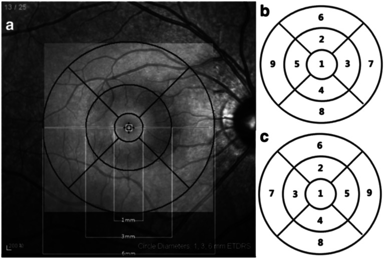
Sixteen eyes of 12 DME patients were included in this study conducted prospectively. All eyes underwent functional and morphologic examination of the macular area using microperimetry and optical coherence tomography (OCT) before and after intravitreal anti-VEGF injection. To determine significant differences between the values, paired t test was used. A correlation between CMT and RS was made using Spearman's test.
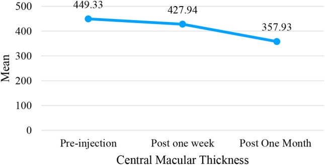
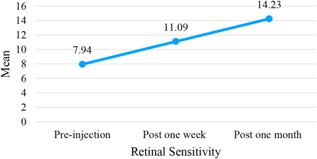
Patients were evaluated at baseline, one week and one month after injection. The central macular thickness (CMT) decreased significantly from 449.33 ± 100.79 μm to 427.94 ± 85.76 μm to 357.93 ± 75.92 μm. The RS improved significantly from 7.94 ± 6.43 dB to 11.09 ± 7.42 dB at one week and to 14.22 ± 7.66 dB at one month after treatment. The CMT was significant negatively correlated to RS (r=-0.259, p = < 0.001), with decay of 0.025 dB for every 1 μm increase of CMT.
Retinal thickening due to DME can be adequately quantified using OCT, while microperimetry can offer information about retinal sensitivity in the exact location. Therefore, microperimetry can be a useful tool in predicting the functional outcome and determining the efficacy of anti-VEGF treatment for DME patients.
评估糖尿病性黄斑水肿(DME)患者玻璃体内注射抗血管内皮生长因子(anti-VEGF)治疗后视网膜敏感度(RS)和视网膜形态的早期反应。
本前瞻性研究纳入了12例DME患者的16只眼。所有眼睛在玻璃体内注射anti-VEGF前后均使用微视野计和光学相干断层扫描(OCT)对黄斑区进行功能和形态学检查。为确定数值之间的显著差异,采用配对t检验。使用Spearman检验分析中心凹视网膜厚度(CMT)与RS之间的相关性。
在注射后基线、1周和1个月时对患者进行评估。中心凹视网膜厚度(CMT)从449.33±100.79μm显著降至427.94±85.76μm,再降至357.93±75.92μm。治疗后1周时RS从7.94±6.43dB显著提高到11.09±7.42dB,1个月时提高到14.22±7.66dB。CMT与RS呈显著负相关(r=-0.259,p<0.001),CMT每增加1μm,RS衰减0.025dB。
OCT可充分量化DME导致的视网膜增厚,而微视野计可提供精确位置的视网膜敏感度信息。因此,微视野计可作为预测DME患者功能转归及确定anti-VEGF治疗疗效的有用工具。