Kamoshita Maki, Shirai Hiroki, Nakamura Hiroko, Kishimoto Tetsuya, Hatanaka Yuki, Mashiko Daisuke, Esashika Katsuhiro, Yang Jingjing, Yamasaki Satoshi, Ogawa Takehiko, Kimura Hiroshi, Ikawa Masahito
Research Institute for Microbial Diseases, Osaka University, Osaka, Japan.
Micro/Nano Technology Center, Tokai University, Kanagawa, Japan.
Sci Rep. 2025 Jan 3;15(1):625. doi: 10.1038/s41598-024-84965-1.
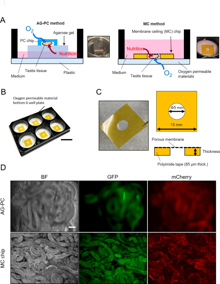
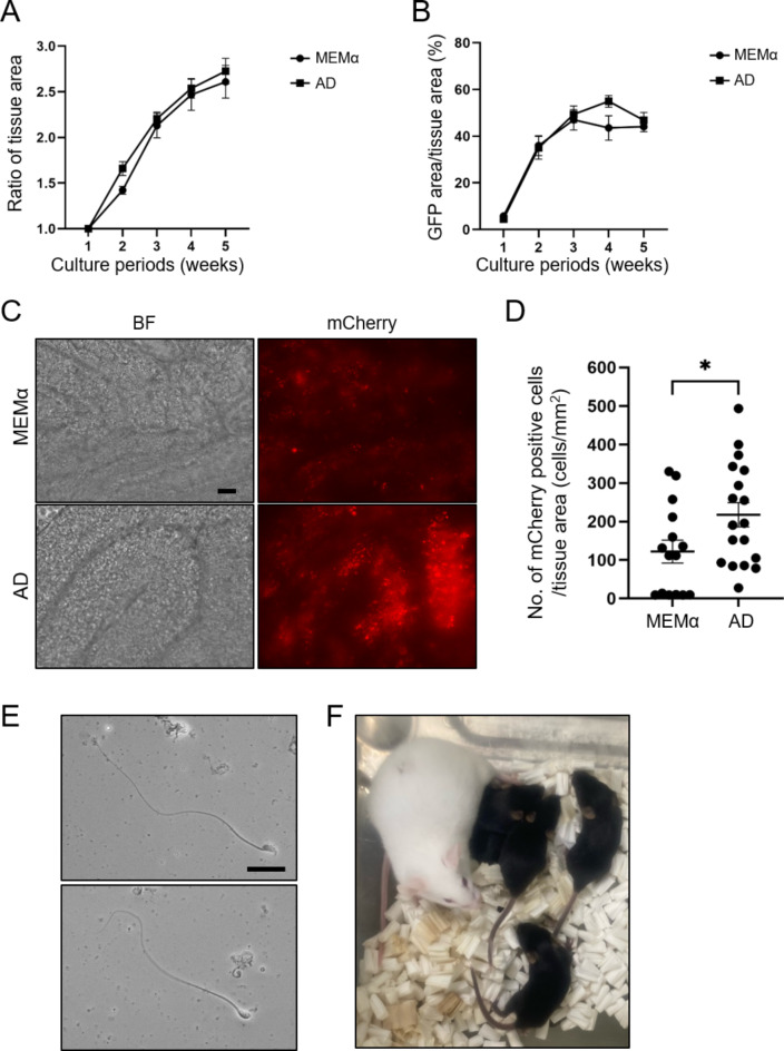
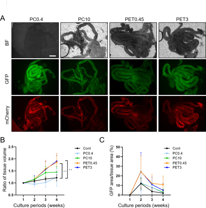
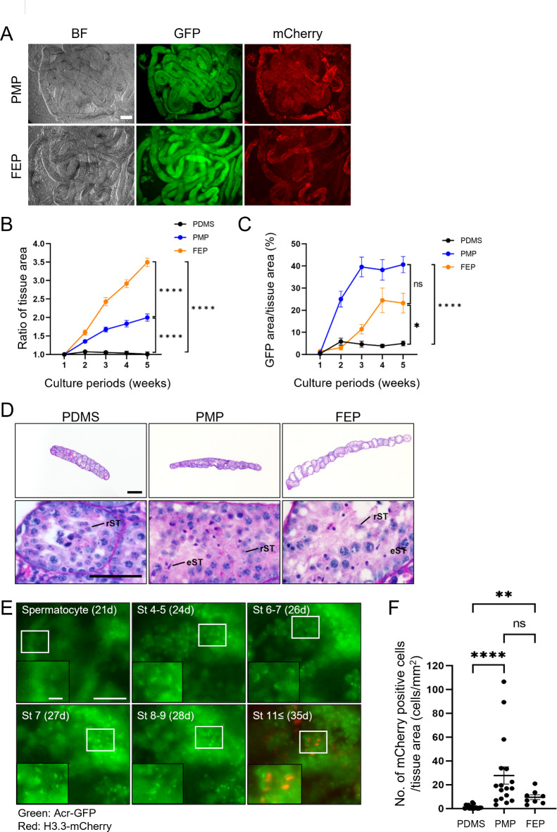
Spermatogenesis is one of the most complex processes of cell differentiation and its failure is a major cause of male infertility. Therefore, a proper model that recapitulates spermatogenesis in vitro has been long sought out for basic and clinical research. Testis organ culture using the gas-liquid interphase method has been shown to support spermatogenesis in mice and rats. However, the conventional method using agarose gel has limitations including medium replacement efficiency and live imaging because agarose absorbs medium and is not transparent. To overcome this issue, we developed a new device using microporous membranes and oxygen-permeable materials. Mouse testes sandwiched between a microporous polyethylene terephthalate (PET) membrane on top and an oxygen-permeable 4-polymethyl-1-pentene polymer (PMP) membrane base maintained spermatogenesis over months. The chamber volume was minimized to 0.1% of the culture medium. Weekly time-lapse live imaging enabled us to observe transgenically fluorescent acrosome and nuclear shape formation throughout spermatogenesis. Finally, we obtained healthy fertile offspring from spermatozoa generated in our system. The device could be used not only for basic research to understand spermatogenesis but also for applied research, such as diagnosing and treating male infertility.
精子发生是细胞分化最复杂的过程之一,其失败是男性不育的主要原因。因此,长期以来一直在寻找一种能够在体外重现精子发生过程的合适模型用于基础和临床研究。使用气液界面法进行睾丸器官培养已被证明可支持小鼠和大鼠的精子发生。然而,使用琼脂糖凝胶的传统方法存在局限性,包括培养基更换效率和实时成像,因为琼脂糖会吸收培养基且不透明。为克服这一问题,我们开发了一种使用微孔膜和透氧材料的新装置。夹在顶部的微孔聚对苯二甲酸乙二酯(PET)膜和底部的透氧4-聚甲基-1-戊烯聚合物(PMP)膜之间的小鼠睾丸在数月内维持了精子发生。腔室体积最小化至培养基的0.1%。每周的延时实时成像使我们能够观察到整个精子发生过程中转基因荧光顶体和细胞核形状的形成。最后,我们从我们系统中产生的精子获得了健康可育的后代。该装置不仅可用于理解精子发生的基础研究,还可用于应用研究,如诊断和治疗男性不育。