Zhang Zhen, Niu Kun, Huang Taoying, Guo Jiali, Xarbat Gongbikai, Gong Xiaoli, Gao Yunke, Liu Feiyang, Cheng Shan, Su Wenting, Yang Fei, Liu Zhaoyuan, Ginhoux Florent, Zhang Ting
Department of Neurobiology, Center of Parkinson Disease Beijing Institute for Brain Disorders, Beijing Key Laboratory on Parkinson Disease, Key Laboratory for Neurodegenerative Disease of the Ministry of Education, Beijing Key Laboratory of Neural Regeneration and Repair, Capital Medical University, Beijing, 100069, China.
Department of Physiology and Pathophysiology, Capital Medical University, Beijing, 100069, China.
NPJ Parkinsons Dis. 2025 Jan 9;11(1):15. doi: 10.1038/s41531-024-00846-4.
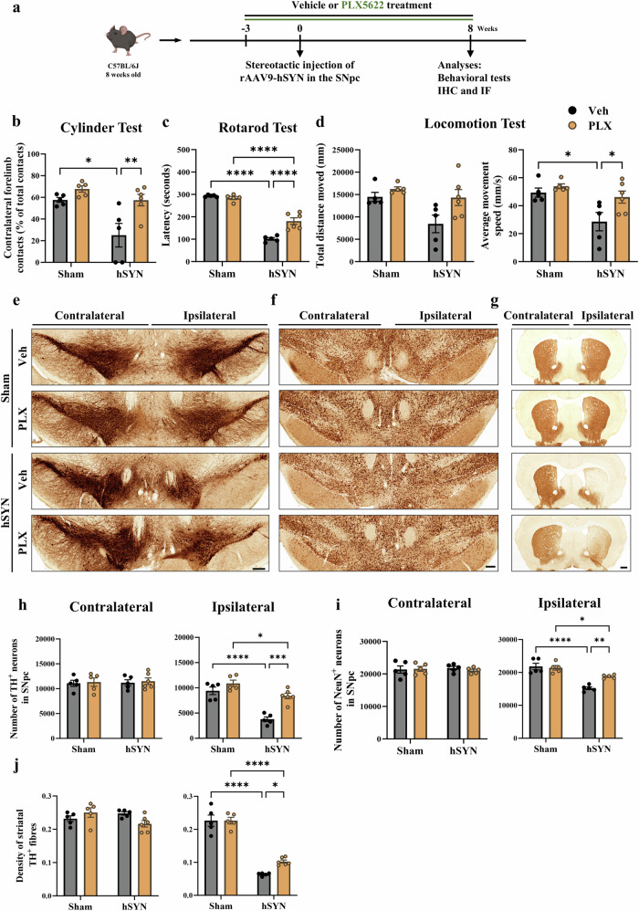
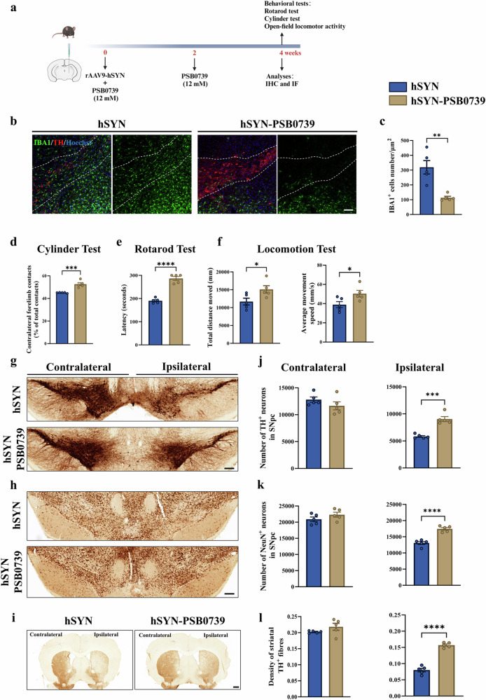
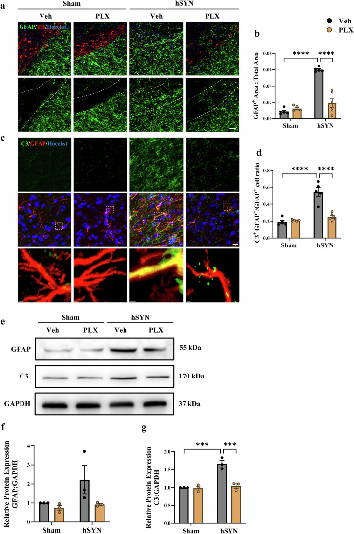
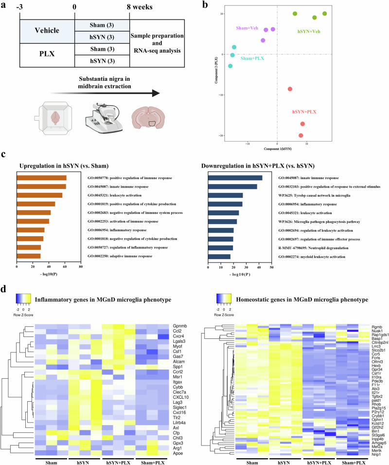
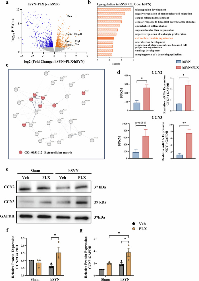
Chronic neuroinflammation with sustained microglial activation occurs in Parkinson's disease (PD), yet the mechanisms and exact contribution of these cells to the neurodegeneration remains poorly understood. In this study, we induced progressive dopaminergic neuron loss in mice via rAAV-hSYN injection to cause the neuronal expression of α-synuclein, which produced neuroinflammation and behavioral alterations. We administered PLX5622, a colony-stimulating factor 1 receptor inhibitor, for 3 weeks prior to rAAV-hSYN injection, maintaining it for 8 weeks to eliminate microglia. This chronic treatment paradigm prevented the development of motor deficits and concomitantly preserved dopaminergic neuron cell and weakened α-synuclein phosphorylation. Gene expression profiles related to extracellular matrix (ECM) remodeling were increased after microglia depletion in PD mice, which were further validated on protein level. We demonstrated that microglia exert adverse effects during α-synuclein-overexpression-induced neuronal lesion formation, and their depletion remodels ECM and aids recovery following insult.
帕金森病(PD)中会出现伴有小胶质细胞持续激活的慢性神经炎症,然而这些细胞对神经退行性变的机制及确切作用仍知之甚少。在本研究中,我们通过注射rAAV-hSYN诱导小鼠多巴胺能神经元进行性丧失,以引发α-突触核蛋白的神经元表达,从而产生神经炎症和行为改变。在注射rAAV-hSYN前3周,我们给予集落刺激因子1受体抑制剂PLX5622,并持续给药8周以清除小胶质细胞。这种慢性治疗模式可预防运动功能障碍的发展,同时保留多巴胺能神经元细胞并减弱α-突触核蛋白磷酸化。在PD小鼠小胶质细胞耗竭后,与细胞外基质(ECM)重塑相关的基因表达谱增加,并在蛋白质水平上得到进一步验证。我们证明,小胶质细胞在α-突触核蛋白过表达诱导的神经元损伤形成过程中发挥不利作用,其耗竭可重塑ECM并有助于损伤后的恢复。