Imaoka Eiki, Nishihori Masahiro, Izumi Takashi, Goto Shunsaku, Araki Yoshio, Yokoyama Kinya, Uda Kenji, Kanamori Fumiaki, Saito Ryuta
Department of Neurosurgery, Nagoya University Graduate School of Medicine, Nagoya, Japan.
Department of Neurosurgery, Japanese Red Cross Aichi Medical Center Nagoya Daini Hospital, Nagoya, Japan.
Nagoya J Med Sci. 2024 Nov;86(4):655-664. doi: 10.18999/nagjms.86.4.655.
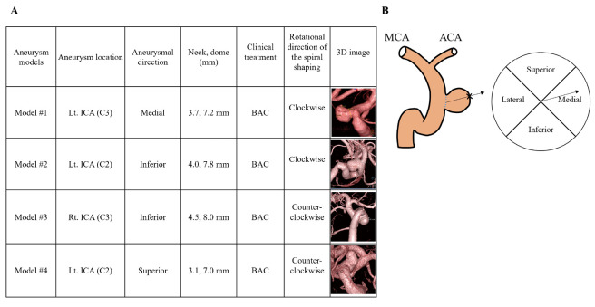
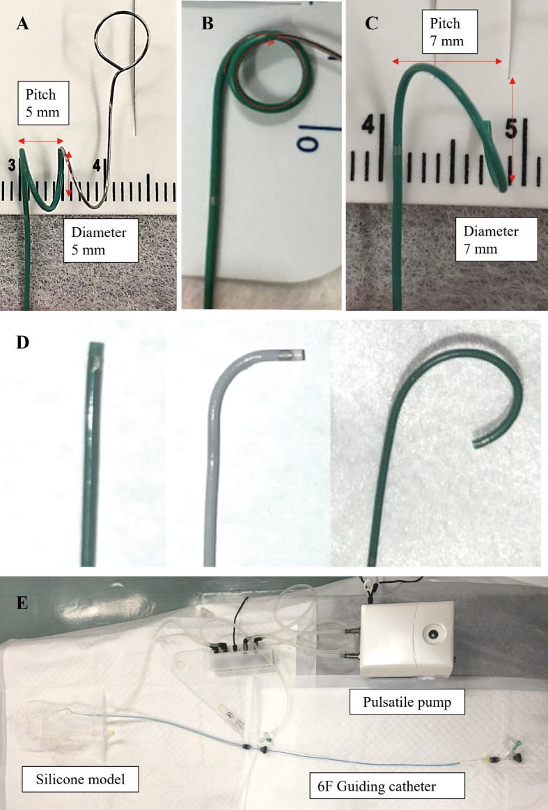
Selecting an appropriate microcatheter tip shape for paraclinoid aneurysms is difficult. Therefore, we devised an original simple and uniform three-dimensional (3D) spiral-shaping method of microcatheter and validated the characteristics and usefulness of this method for coil embolization of paraclinoid aneurysms using patient-specific silicone models. These silicone models were produced based on clinical data from four patients with four paraclinoid aneurysms that underwent endovascular treatment using the 3D spiral-shaping method. These models were classified into four types: superior, medial, inferior, and lateral corresponding to the aneurysm protrusion and locations (C3 or C2 segments by Fisher's classification). Employing a pulsatile pump setup, two operators assessed the following items: navigation methods (pull and wire guiding), catheterization times, microcatheter tip position in the aneurysm, and the feasibility of inserting a framing coil by simple technique compared with three other shapes (straight, 90, pigtail). Three-dimensional spiral-shaped microcatheter could be placed in the medial and inferior type models of C3 segments and superior type model of C2 segment by the pullback method. Catheterization times using a 3D spiral-shaped catheter were significantly shorter than other shaped ones in the superior type models. No significant difference was found in another silicone model. Three-dimensional spiral- and pigtail-shaped catheters tended to position the tip at the center of the aneurysm. In conclusion, 3D spiral-shaped microcatheter was especially effective for the superior projected aneurysm at the C2 segment. The 3D spiral-shaping method can provide easy and secure navigation of the microcatheter into the paraclinoid aneurysms, ensuring optimal positioning for coil insertion.
为海绵窦旁动脉瘤选择合适的微导管尖端形状很困难。因此,我们设计了一种原创的、简单且统一的微导管三维(3D)螺旋塑形方法,并使用患者特异性硅胶模型验证了该方法在海绵窦旁动脉瘤弹簧圈栓塞中的特性和实用性。这些硅胶模型是根据4例患有4个海绵窦旁动脉瘤的患者的临床数据制作的,这些患者接受了使用3D螺旋塑形方法的血管内治疗。这些模型分为四种类型:上、中、下和外侧,分别对应动脉瘤的突出部位和位置(根据Fisher分类法为C3或C2段)。采用脉动泵装置,两名操作人员评估了以下项目:导航方法(回撤和导丝引导)、插管时间、微导管尖端在动脉瘤内的位置,以及与其他三种形状(直形、90°、猪尾形)相比,通过简单技术插入成篮弹簧圈的可行性。通过回撤法,三维螺旋形微导管可放置在C3段的内侧和下侧型模型以及C2段的上侧型模型中。在上侧型模型中,使用3D螺旋形导管的插管时间明显短于其他形状的导管。在另一个硅胶模型中未发现显著差异。三维螺旋形和猪尾形导管倾向于将尖端置于动脉瘤中心。总之,3D螺旋形微导管对C2段上向突出的动脉瘤特别有效。3D螺旋塑形方法可为微导管轻松、安全地进入海绵窦旁动脉瘤提供导航,确保弹簧圈插入的最佳定位。