Holländer Sebastian, von Heesen Maximilian, Gäbelein Gereon, Mercier Julie, Laschke Matthias W, Menger Michael D, Glanemann Matthias, Spiliotis Antonios E
Department of General Surgery, Vascular-, Visceral- and Pediatric Surgery, Saarland University Medical Center, 66421, Homburg, Germany.
Department of General- and Visceral Surgery, University Hospital Göttingen, 37075, Göttingen, Germany.
Sci Rep. 2025 Jan 22;15(1):2753. doi: 10.1038/s41598-025-87135-z.
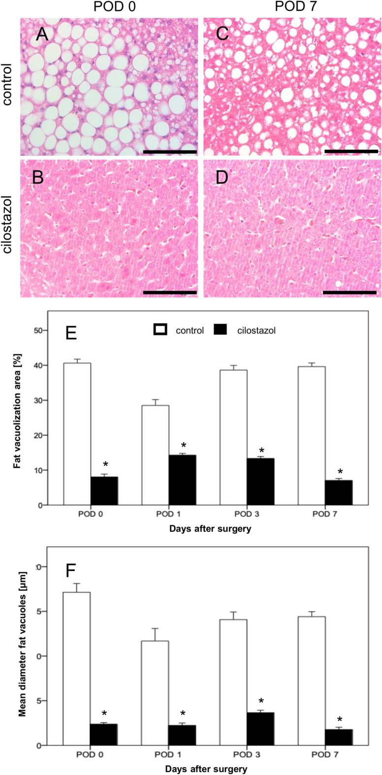
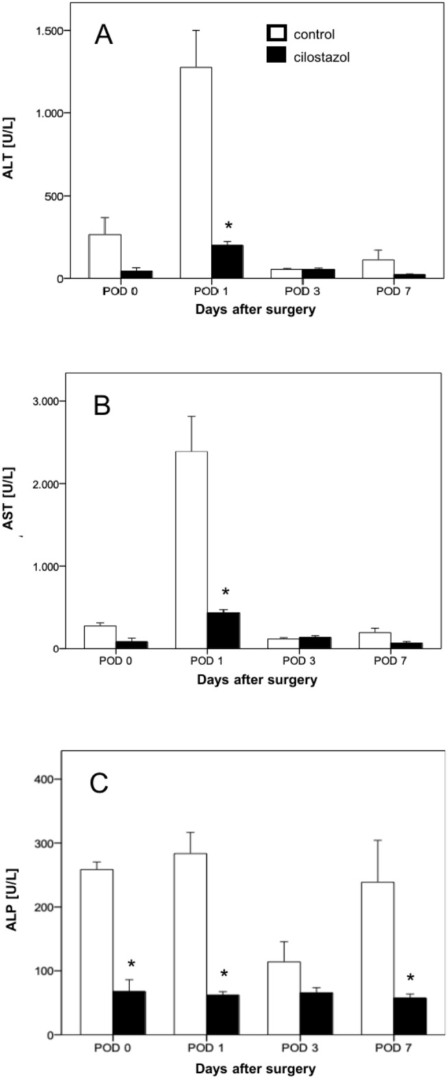
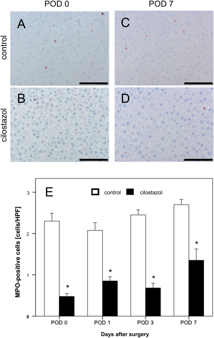
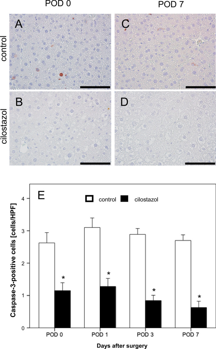
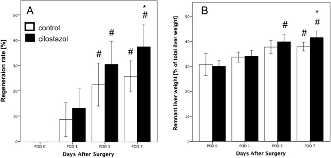
Cilostazol has previously been shown to reduce liver steatosis and enhance hepatic perfusion. We investigated the effects of cilostazol after major hepatectomy in a steatotic rat model. Six weeks prior to surgery, Sprague-Dawley rats were fed with a high-fructose diet. The treatment group received daily 5 mg/kg cilostazol. Seven days following the cilostazol treatment, all animals underwent 70% liver resection (PHX). Analysis of hepatic blood flow and microcirculation and immunohistochemical examinations were conducted 30 min after PHX (postoperative day [POD] 0) as well as on POD 1, POD 3 and POD 7. The weight of cilostazol-treated animals was significantly reduced compared to untreated controls after completion of the 6-week high-FRC diet. Furthermore, 41% macrovesicular steatosis was found in the control group compared to 8% in the cilostazol group. Hepatic arterial and portal venous perfusion were increased in the cilostazol group on POD 7. Lower liver enzyme release was found postoperatively in cilostazol-treated animals. Moreover, apoptosis and neutrophil infiltration were reduced after cilostazol treatment. Proliferation of hepatocytes and liver regeneration after PHX were significantly increased in the cilostazol group. Consequently, cilostazol should be evaluated as a novel strategy to reduce the rate of liver failure after PHX in steatotic liver.
西洛他唑先前已被证明可减轻肝脂肪变性并增强肝脏灌注。我们在脂肪变性大鼠模型中研究了西洛他唑在大肝切除术后的作用。手术前六周,给Sprague-Dawley大鼠喂食高果糖饮食。治疗组每天接受5mg/kg西洛他唑。西洛他唑治疗七天后,所有动物均接受70%肝切除术(PHX)。在PHX后30分钟(术后第0天)以及术后第1天、第3天和第7天进行肝血流和微循环分析以及免疫组化检查。在完成六周高果糖饮食后,与未治疗的对照组相比,西洛他唑治疗动物的体重显著降低。此外,对照组中发现41%的大泡性脂肪变性,而西洛他唑组为8%。在术后第7天,西洛他唑组的肝动脉和门静脉灌注增加。西洛他唑治疗的动物术后肝酶释放较低。此外,西洛他唑治疗后细胞凋亡和中性粒细胞浸润减少。西洛他唑组PHX后肝细胞增殖和肝再生显著增加。因此,应评估西洛他唑作为一种降低脂肪变性肝脏PHX后肝衰竭发生率的新策略。