Moradicheghamahi Jafar, Fortuny Gerard, López Josep M, Puigjaner Dolors, Herrero Joan, Azeli Youcef
Departament d'Enginyeria Informàtica i Matemàtiques, Universitat Rovira i Virgili, 43007 Tarragona, Spain.
Departament d'Enginyeria Química, Universitat Rovira i Virgili, 43007 Tarragona, Spain.
Resusc Plus. 2025 Feb 18;22:100910. doi: 10.1016/j.resplu.2025.100910. eCollection 2025 Mar.
Basic science research in cardiopulmonary resuscitation (CPR) is limited by challenges in obtaining haemodynamic data from models that simulate physiological processes. In this study, we assessed the morphology of the heart and lungs and calculated the ejection fractions of cardiac chambers during CPR using a virtual simulation.
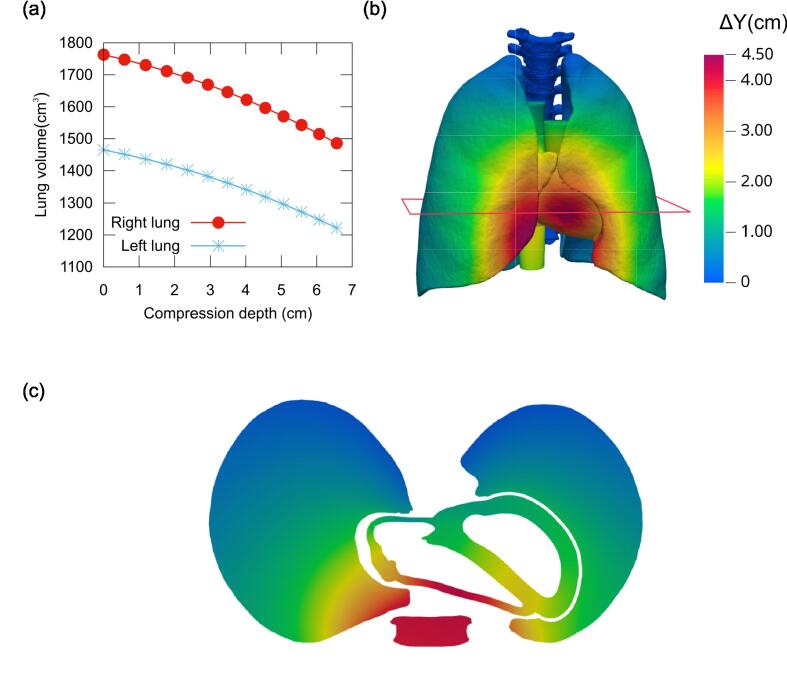
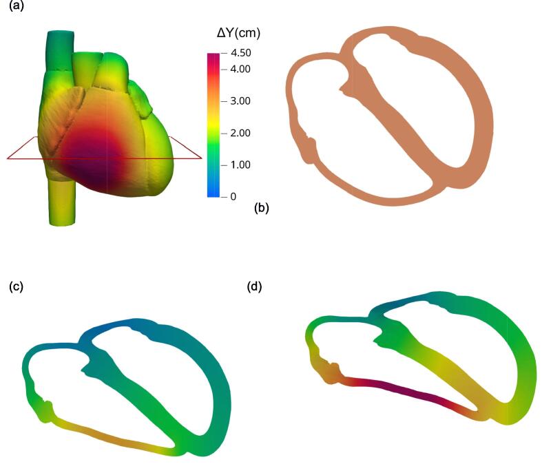
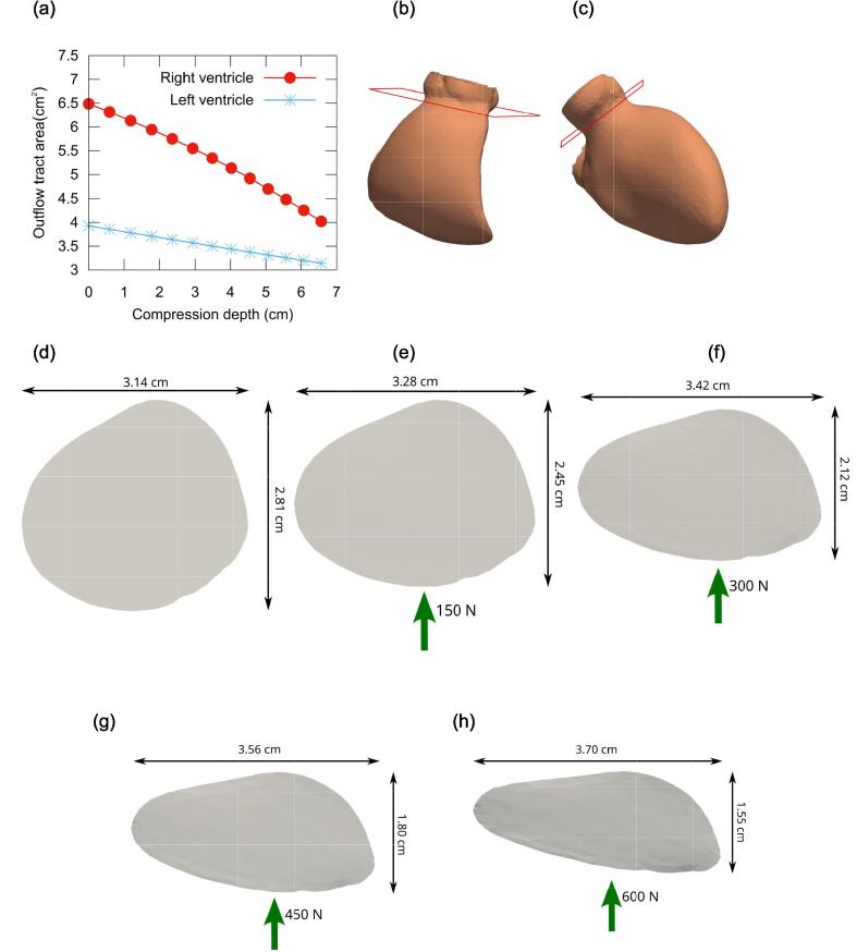
A finite element model of a complete thorax, including internal organs, thoracic rib cage, spine, musculature, and a generic material representing soft tissues, was constructed from magentic resonance images of a man. Twelve chest compression simulations were performed with forces ranging from = 50 to 600 N. During compression, lung and heart volumes were assessed, and the ejection fraction of each cardiac chamber was calculated.
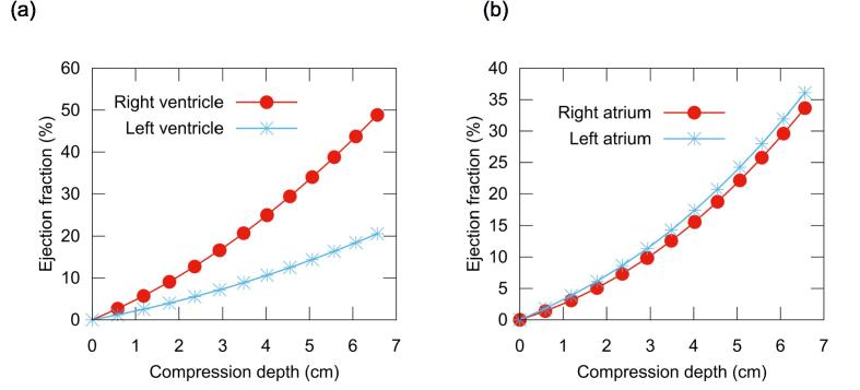
In our numerical simulations a compression depth of 5.06 cm was reached with a force of 450 N. At this depth, the right and left ventricular ejection fractions were 34.0% and 14.4%, respectively, while the right and left atrial ejection fractions were 22.1% and 24.2%, respectively. The cross-sectional area of the outflow tract decreased by 27.5% and 15.6% in the right and left ventricles, respectively. Lung volumes decreased by 193 cm and 169 cm in the right and left lungs, respectively, representing 11.2% of the total lung volume.
The right ventricle exhibited the highest ejection fraction among the cardiac chambers, and the left atrium showed a higher ejection fraction than the left ventricle during CPR.
心肺复苏(CPR)的基础科学研究受到从模拟生理过程的模型获取血流动力学数据方面挑战的限制。在本研究中,我们使用虚拟模拟评估了心肺复苏期间心脏和肺部的形态,并计算了心腔的射血分数。
根据一名男性的磁共振图像构建了一个完整胸部的有限元模型,包括内部器官、胸廓肋骨、脊柱、肌肉组织以及代表软组织的通用材料。使用50至600N的力进行了12次胸外按压模拟。在按压过程中,评估肺和心脏体积,并计算每个心腔的射血分数。
在我们的数值模拟中,450N的力可使按压深度达到5.06cm。在此深度时,右心室和左心室的射血分数分别为34.0%和14.4%,而右心房和左心房的射血分数分别为22.1%和24.2%。右心室和左心室流出道的横截面积分别减少了27.5%和15.6%。右肺和左肺的肺体积分别减少了193cm³和169cm³,占总肺体积的11.2%。
在心腔中,右心室的射血分数最高,并且在心肺复苏期间左心房的射血分数高于左心室。