Shahin Hady, Steinvall Ingrid, Sjöberg Folke, Elmasry Moustafa, El-Serafi Ahmed
Department of Hand Surgery, Plastic Surgery and Burns, Linköping University, Linköping, Sweden.
The Department of Biomedical and Clinical Sciences, Linköping University, Linköping, Sweden.
Front Bioeng Biotechnol. 2025 Mar 20;13:1547044. doi: 10.3389/fbioe.2025.1547044. eCollection 2025.
Human keratinocytes require relatively long propagation time which impedes their availability as autologous cell transplantation within a clinically reasonable timeframe. There is an unmet need for efficient xeno-free cell expansion approaches to propagate human keratinocytes as regenerative therapy.
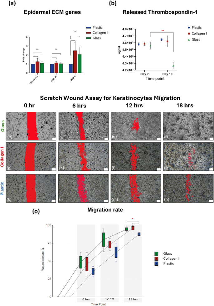
Primary human keratinocytes and HaCaT cells were cultured on glass, plastic, and animal-derived collagen I matrix for 10 days. Proliferation, migration, DNA methylation, as well as gene and protein expression were assessed to characterize the effect of the tested culture substrates on keratinocytes at the molecular and functional levels.
Keratinocytes cultured on glass exhibited faster proliferation, global DNA demethylation and upregulation of epidermal differentiation markers. Scratch wound assay revealed that keratinocytes cultured on glass demonstrated enhanced cell migration compared to those on plastic or collagen I. Multiplex immunoassays identified temporal and substrate-dependent variations in a panel of keratinocyte-specific secreted factors, encompassing immunomodulatory cytokines, growth factors, and angiogenic factors.
Glass, as a culture substrate, promotes epidermal differentiation and enhances keratinocyte migration. The latter is a critical factor in re-epithelialization and wound healing. Functional properties suggest that glass may optimize the inflammatory response and promote efficient wound repair, making it a promising candidate for the short-term expansion of keratinocytes for transplantation purposes. Further validation is required to definitively establish the efficacy of keratinocytes cultured on glass for clinical applications.
人角质形成细胞需要相对较长的增殖时间,这阻碍了它们在临床合理时间范围内作为自体细胞移植的可用性。对于有效的无动物源细胞扩增方法来扩增人角质形成细胞用于再生治疗,存在未满足的需求。
将原代人角质形成细胞和HaCaT细胞在玻璃、塑料和动物源I型胶原基质上培养10天。评估增殖、迁移、DNA甲基化以及基因和蛋白质表达,以在分子和功能水平上表征测试的培养底物对角质形成细胞的影响。
在玻璃上培养的角质形成细胞表现出更快的增殖、全基因组DNA去甲基化以及表皮分化标志物的上调。划痕伤口试验表明,与在塑料或I型胶原上培养的角质形成细胞相比,在玻璃上培养的角质形成细胞表现出增强的细胞迁移。多重免疫测定确定了一组角质形成细胞特异性分泌因子的时间和底物依赖性变化,包括免疫调节细胞因子、生长因子和血管生成因子。
玻璃作为一种培养底物,可促进表皮分化并增强角质形成细胞迁移。后者是再上皮化和伤口愈合的关键因素。功能特性表明,玻璃可优化炎症反应并促进有效的伤口修复,使其成为用于移植目的的角质形成细胞短期扩增的有希望的候选者。需要进一步验证以最终确定在玻璃上培养的角质形成细胞用于临床应用的疗效。