Gaffuri Michele, Battilocchi Ludovica, Lazzeroni Matteo, Pignataro Lorenzo, Capaccio Pasquale
Department of Otolaryngology and Head and Neck Surgery, Fondazione IRCCS Ca' Granda Ospedale Maggiore Policlinico, 20122 Milan, Italy.
Department of Clinical Sciences and Community Health, Dipartimento di Eccellenza 2023-2027, University of Milan, 20122 Milan, Italy.
J Clin Med. 2025 Mar 28;14(7):2338. doi: 10.3390/jcm14072338.
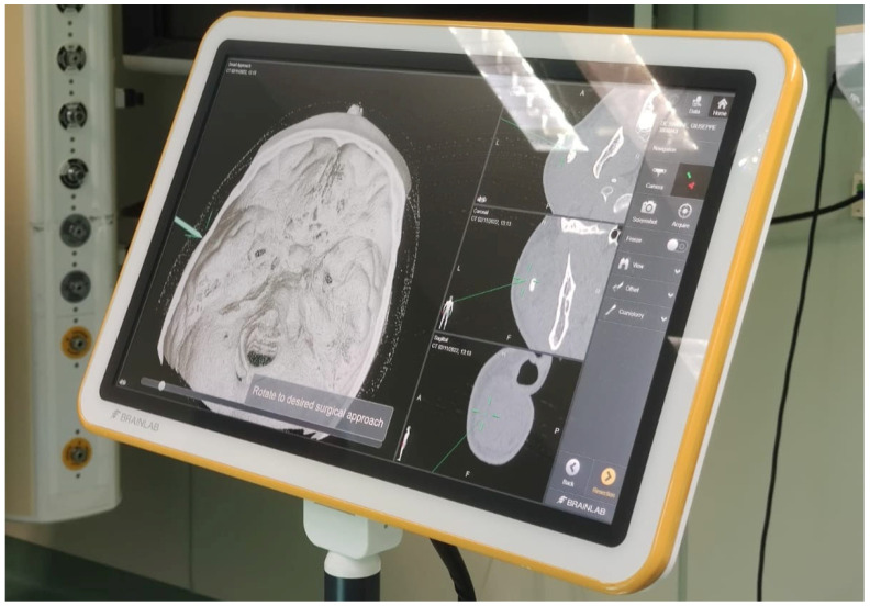
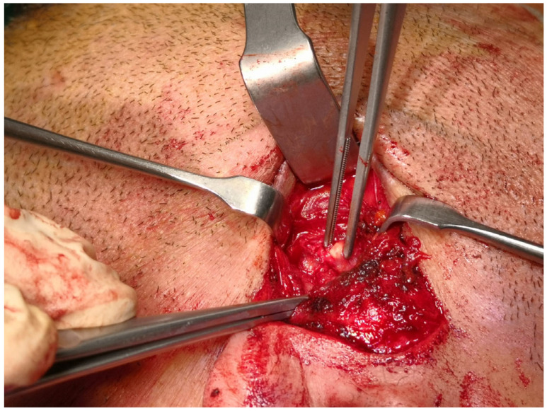
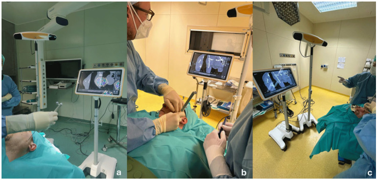
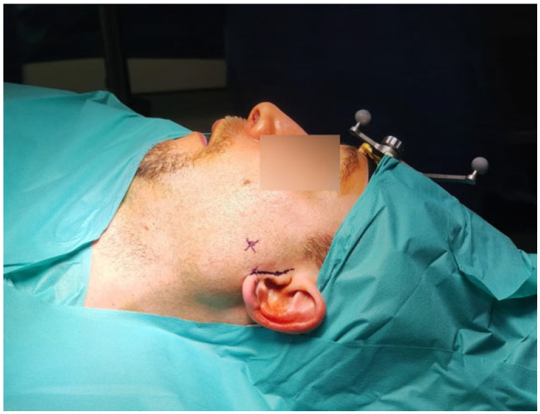
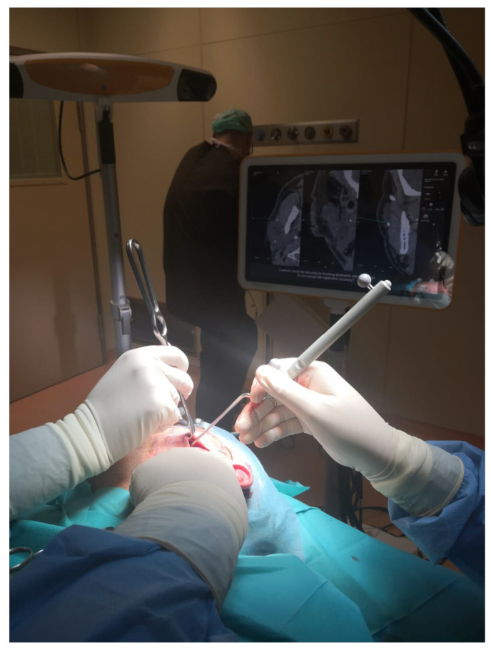
The failure rate of minimally invasive surgical approaches to parotid stones is about 10%, primarily due to the presence of large, impacted, or unpalpable deep stones. When stones are palpable and exceed 7 mm in size, a combined transfacial and sialendoscopic approach offers a safe and effective surgical option, while unpalpable and impacted stones located in the parenchyma, not visible or accessible through sialendoscopy, can be treated with a CT-guided transfacial approach. Twenty-two patients (three females, mean age 53 years, range 32-73 years) underwent CT navigation-assisted transfacial removal of unpalpable and impacted parotid stones at the Department of Otolaryngology and Head and Neck Surgery of Fondazione IRCCS Ca' Granda Ospedale Maggiore Policlinico of Milan between 2017 and 2024. The mean size of the stones was 7.4 mm (range 4-14 mm), while the mean depth of stones, calculated as the distance from the skin surface, was 8.7 mm (range 4-14 mm). Stones were removed successfully in all but five patients (77% success rate). Failure of the procedure was significantly associated ( < 0.05) with the depth of the stone (>12 mm); in all these cases, patients were treated immediately by means of traditional parotidectomy. The CT-navigation-assisted transfacial approach can be considered a safe, reliable, and efficacious option for the treatment of difficult unpalpable parotid stones, impacted and deeply located in the gland parenchyma. Stones deeper than 10 mm can be more effectively treated by means of traditional parotidectomy if extracorporeal lithotripsy is not available.
腮腺结石微创外科手术方法的失败率约为10%,主要原因是存在较大、嵌顿或无法触及的深部结石。当结石可触及且大小超过7mm时,经面部联合唾液腺内镜手术是一种安全有效的手术选择,而位于实质内、无法触及且嵌顿的结石,唾液腺内镜不可见或无法触及,则可采用CT引导下经面部手术治疗。2017年至2024年期间,22例患者(3例女性,平均年龄53岁,范围32 - 73岁)在米兰 Fondazione IRCCS Ca' Granda Ospedale Maggiore Policlinico的耳鼻喉科和头颈外科接受了CT导航辅助经面部切除无法触及且嵌顿的腮腺结石手术。结石的平均大小为7.4mm(范围4 - 14mm),而结石的平均深度(从皮肤表面计算)为8.7mm(范围4 - 14mm)。除5例患者外,所有患者的结石均成功取出(成功率77%)。手术失败与结石深度(>12mm)显著相关(<0.05);在所有这些病例中,患者立即接受了传统腮腺切除术治疗。CT导航辅助经面部手术可被认为是治疗位于腮腺实质内难以触及、嵌顿且深部的腮腺结石的一种安全、可靠且有效的选择。如果没有体外碎石术,深度超过10mm的结石通过传统腮腺切除术可得到更有效的治疗。