Streller Matthias, Michlíková Soňa, Ciecior Willy, Lönnecke Katharina, Kunz-Schughart Leoni A, Lange Steffen, Voss-Böhme Anja
DataMedAssist Group, HTW Dresden-University of Applied Sciences, 01069 Dresden, Germany.
Faculty of Informatics/Mathematics, HTW Dresden-University of Applied Sciences, 01069 Dresden, Germany.
Gigascience. 2025 Jan 6;14. doi: 10.1093/gigascience/giaf027.
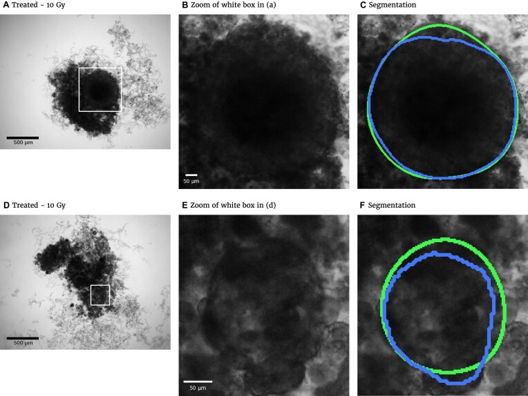
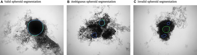
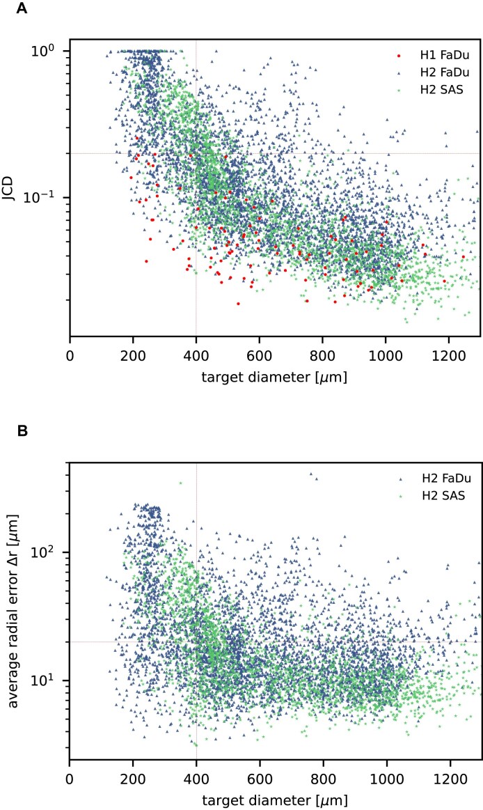
Multicellular tumor spheroids (MCTS) are advanced cell culture systems for assessing the impact of combinatorial radio(chemo)therapy as they exhibit therapeutically relevant in vivo-like characteristics from 3-dimensional cell-cell and cell-matrix interactions to radial pathophysiological gradients. State-of-the-art assays quantify long-term curative endpoints based on collected brightfield image time series from large treated spheroid populations per irradiation dose and treatment arm. This analyses require laborious spheroid segmentation of up to 100,000 images per treatment arm to extract relevant structural information from the images (e.g., diameter, area, volume, and circularity). While several image analysis algorithms are available for spheroid segmentation, they all focus on compact MCTS with a clearly distinguishable outer rim throughout growth. However, they often fail for the common case of treated MCTS, which may partly be detached and destroyed and are usually obscured by dead cell debris.
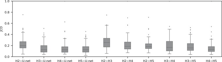
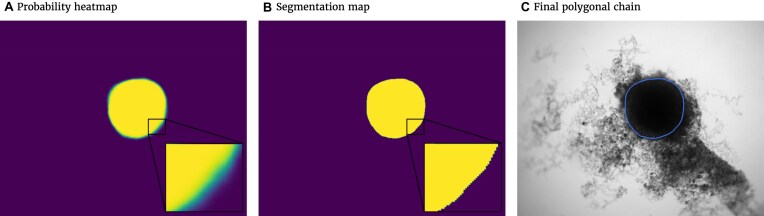
To address these issues, we successfully train 2 fully convolutional networks, UNet and HRNet, and optimize their hyperparameters to develop an automatic segmentation for both untreated and treated MCTS. We extensively test the automatic segmentation on larger, independent datasets and observe high accuracy for most images with Jaccard indices around 90%. For cases with lower accuracy, we demonstrate that the deviation is comparable to the interobserver variability. We also test against previously published datasets and spheroid segmentations.
The developed automatic segmentation can not only be used directly but also integrated into existing spheroid analysis pipelines and tools. This facilitates the analysis of 3-dimensional spheroid assay experiments and contributes to the reproducibility and standardization of this preclinical in vitro model.
多细胞肿瘤球体(MCTS)是用于评估联合放(化)疗影响的先进细胞培养系统,因为它们展现出从三维细胞 - 细胞和细胞 - 基质相互作用到径向病理生理梯度等与治疗相关的体内样特征。目前最先进的检测方法基于每个照射剂量和治疗组的大量处理后球体群体收集的明场图像时间序列来量化长期治愈终点。这种分析需要对每个治疗组多达100,000张图像进行费力的球体分割,以从图像中提取相关结构信息(例如直径、面积、体积和圆度)。虽然有几种图像分析算法可用于球体分割,但它们都专注于在整个生长过程中外缘清晰可辨的紧凑MCTS。然而,对于处理后的MCTS这种常见情况,它们常常失败,处理后的MCTS可能部分 detached 和被破坏,并且通常被死细胞碎片遮挡。
为了解决这些问题,我们成功训练了2个全卷积网络,即UNet和HRNet,并优化了它们的超参数,以开发针对未处理和处理后的MCTS的自动分割方法。我们在更大的独立数据集上广泛测试了自动分割方法,并观察到大多数图像的准确率很高,杰卡德指数约为90%。对于准确率较低的情况,我们证明偏差与观察者间的变异性相当。我们还与先前发表的数据集和球体分割方法进行了对比测试。
所开发的自动分割方法不仅可以直接使用,还可以集成到现有的球体分析管道和工具中。这有助于对三维球体检测实验进行分析,并有助于提高这种临床前体外模型的可重复性和标准化。