• 文献检索
  • 文档翻译
  • 深度研究
  • 学术资讯
  • Suppr Zotero 插件Zotero 插件
  • 邀请有礼
  • 套餐&价格
  • 历史记录
应用&插件
Suppr Zotero 插件Zotero 插件浏览器插件Mac 客户端Windows 客户端微信小程序
定价
高级版会员购买积分包购买API积分包
服务
文献检索文档翻译深度研究API 文档MCP 服务
关于我们
关于 Suppr公司介绍联系我们用户协议隐私条款
关注我们

Suppr 超能文献

核心技术专利:CN118964589B侵权必究
粤ICP备2023148730 号-1Suppr @ 2026

文献检索

告别复杂PubMed语法,用中文像聊天一样搜索,搜遍4000万医学文献。AI智能推荐,让科研检索更轻松。

立即免费搜索

文件翻译

保留排版,准确专业,支持PDF/Word/PPT等文件格式,支持 12+语言互译。

免费翻译文档

深度研究

AI帮你快速写综述,25分钟生成高质量综述,智能提取关键信息,辅助科研写作。

立即免费体验

SHP2介导的活性氧激活在骨关节炎中诱导软骨细胞副凋亡,并被低强度脉冲超声减弱。

SHP2-mediated ROS activation induces chondrocyte paraptosis in osteoarthritis and is attenuated by low-intensity pulsed ultrasound.

作者信息

Hou Wenjie, Shang Xingru, Hao Xiaoxia, Pan Chunran, Zheng Zehang, Zhang Yiwen, Deng Xiaofeng, Chi Ruimin, Liu Jiawei, Guo Fengjing, Sun Kai, Xu Tao

机构信息

Department of Rehabilitation, Tongji Hospital, Tongji Medical College, Huazhong University of Science and Technology, Wuhan, 430022, China.

Department of Rehabilitation Medicine,Key Laboratory of Physical Medicine and Precision Rehabilitation of Chongqing Municipal Health Commission, The First Affiliated Hospital of Chongqing Medical University, No.1 Youyi Road, Yuzhong District, Chongqing, 400016, China.

出版信息

J Orthop Translat. 2025 Apr 25;52:233-248. doi: 10.1016/j.jot.2025.04.005. eCollection 2025 May.

DOI:10.1016/j.jot.2025.04.005
PMID:40337549
原文链接:https://pmc.ncbi.nlm.nih.gov/articles/PMC12056802/
Abstract

BACKGROUND

Paraptosis is a novel form of programmed cell death, generally caused by disrupted proteostasis or alterations of redox homeostasis. However, its impact and underlying mechanisms on the pathology of osteoarthritis (OA) are still unclear. This study aimed to investigate the role and regulatory mechanism of SHP2 in chondrocyte paraptosis and the effects influenced by low-intensity pulsed ultrasound (LIPUS).

METHODS

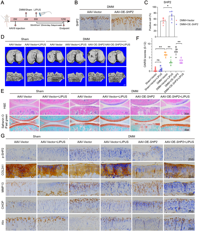
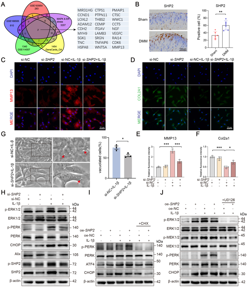
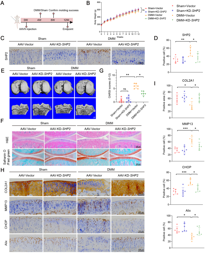
SHP2, a MAPK upstream intermediary, has been identified as one of the critical targets of IL-1β-induced paraptosis in the GEO and GeneCard databases. The expression of SHP2 in chondrocytes was regulated by either siRNA knockdown or plasmid overexpression. Additionally, adeno-associated viruses were injected into the knee joints of rats to explore whether SHP2 plays a role in the development of OA. The impact of LIPUS on paraptosis and OA was examined in IL-1β-induced chondrocytes and a post-traumatic OA model, with SHP2 regulation assessed at both cellular and animal levels.

RESULTS

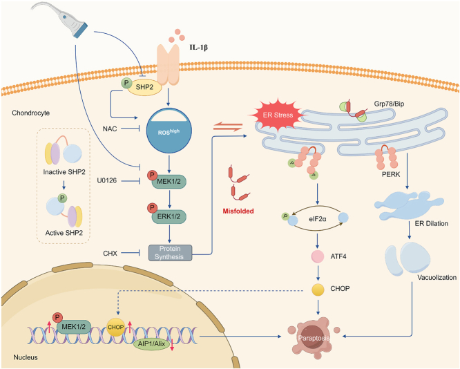
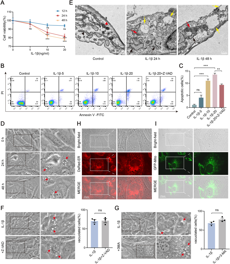
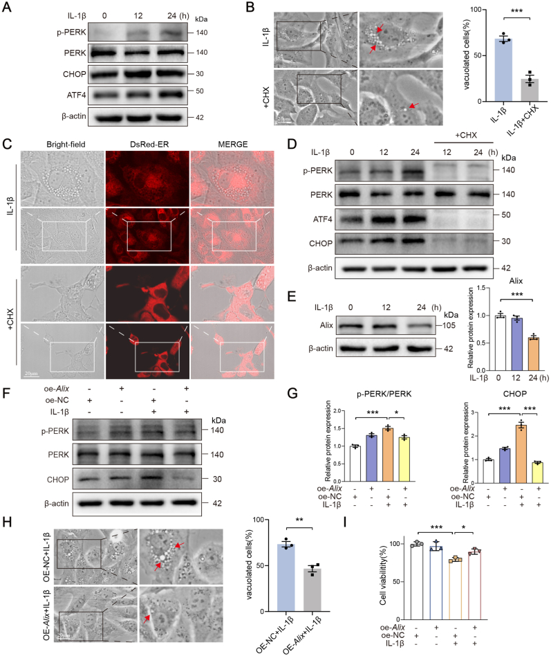
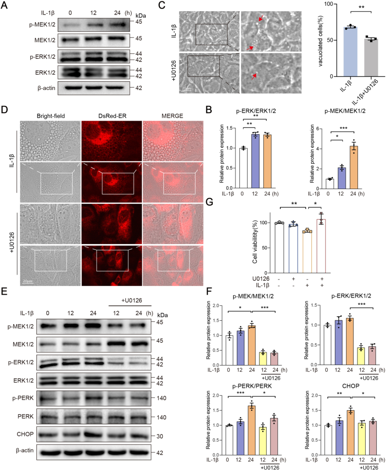
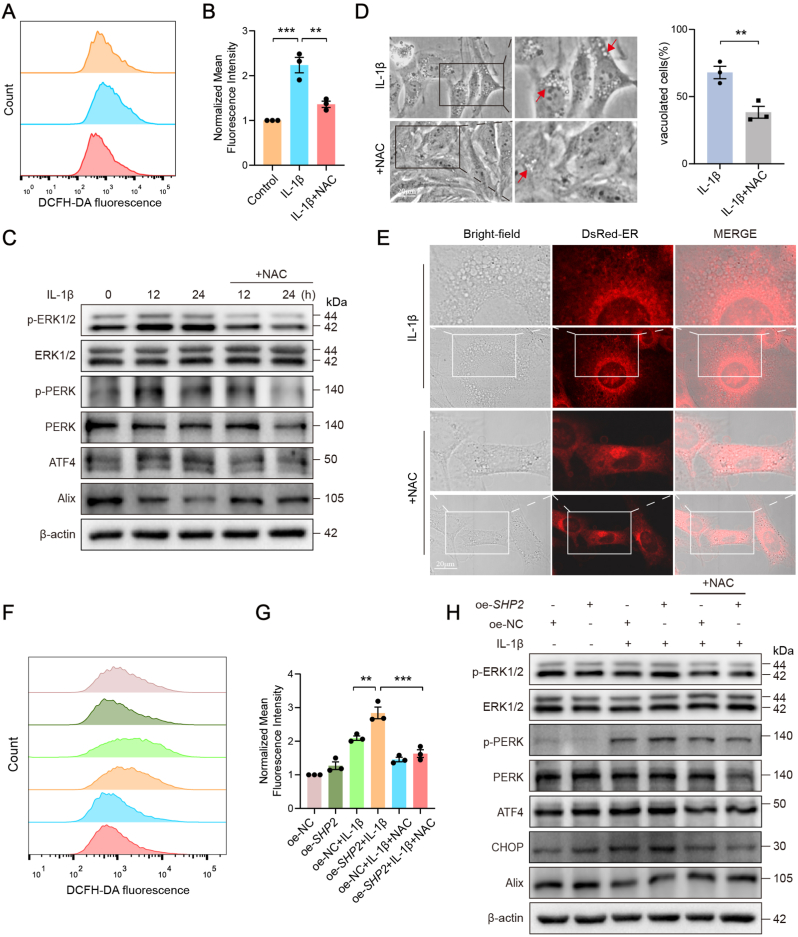
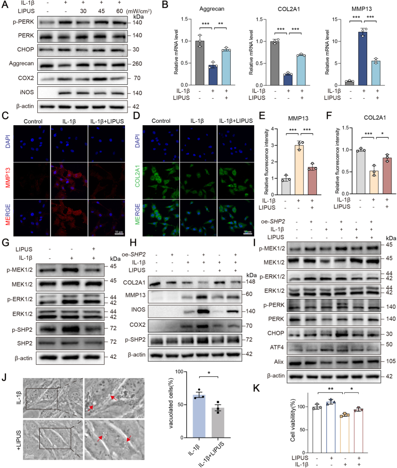
An increase in cellular reactive oxygen species (ROS) caused by IL-1β halts the growth of chondrocytes and induces paraptosis in the chondrocytes. IL-1β-induced paraptosis, manifested as endoplasmic reticulum (ER)-derived vacuolization, was mediated by ROS-mediated ER stress and MAPK activation. SHP2 facilitates ROS production, thereby exacerbating the chondrocytes paraptosis. SHP2 knockdown and ROS inhibition effectively reduced this process and significantly mitigated inflammation and cartilage degeneration. Furthermore, we discovered that LIPUS delayed OA progression by inhibiting the activation of the MAPK pathway, ER stress, and ER-derived vacuoles in chondrocytes, all of which play critical roles in paraptosis, through the downregulation of SHP2 expression. Results on animals showed that LIPUS inhibited cartilage degeneration and alleviated OA progression.

CONCLUSION

SHP2 exacerbates IL-1β-induced oxidative stress and the subsequent paraptosis in chondrocytes, promoting OA progression. LIPUS mitigates paraptosis by modulating SHP2, which in turn slows OA progression.

THE TRANSLATIONAL POTENTIAL OF THIS ARTICLE

This study indicates that a novel SHP2-mediated cell death mechanism, paraptosis, plays a role in post-traumatic OA progression. LIPUS helps maintain cartilage-subchondral bone unit integrity by targeting SHP2 inhibition. SHP2 emerges as a potential therapeutic target, while LIPUS provides a promising non-invasive approach for treating trauma-related OA.

摘要

背景

副凋亡是一种新型程序性细胞死亡形式,通常由蛋白稳态破坏或氧化还原稳态改变引起。然而,其对骨关节炎(OA)病理的影响及潜在机制仍不清楚。本研究旨在探讨SHP2在软骨细胞副凋亡中的作用及调控机制,以及低强度脉冲超声(LIPUS)对其的影响。

方法

在GEO和GeneCard数据库中,已将丝裂原活化蛋白激酶(MAPK)上游中介分子SHP2鉴定为白细胞介素-1β(IL-1β)诱导的副凋亡关键靶点之一。通过小干扰RNA(siRNA)敲低或质粒过表达来调节软骨细胞中SHP2的表达。此外,将腺相关病毒注射到大鼠膝关节中,以探究SHP2是否在OA发展中起作用。在IL-1β诱导的软骨细胞和创伤后OA模型中,检测LIPUS对副凋亡和OA的影响,并在细胞和动物水平评估SHP2的调节情况。

结果

IL-1β引起的细胞活性氧(ROS)增加会阻止软骨细胞生长并诱导软骨细胞发生副凋亡。IL-1β诱导的副凋亡表现为内质网(ER)衍生的空泡化,由ROS介导的内质网应激和MAPK激活介导。SHP2促进ROS产生,从而加剧软骨细胞副凋亡。SHP2敲低和ROS抑制有效减少了这一过程,并显著减轻了炎症和软骨退变。此外,我们发现LIPUS通过下调SHP2表达,抑制MAPK途径激活、内质网应激以及软骨细胞中内质网衍生的空泡,从而延缓OA进展,而这些在内质网应激中均起关键作用。动物实验结果表明,LIPUS抑制了软骨退变并减轻了OA进展。

结论

SHP2加剧IL-1β诱导的氧化应激及随后的软骨细胞副凋亡,促进OA进展。LIPUS通过调节SHP2减轻副凋亡,进而减缓OA进展。

本文的转化潜力

本研究表明,一种新的由SHP2介导的细胞死亡机制——副凋亡,在创伤后OA进展中起作用。LIPUS通过靶向抑制SHP2来帮助维持软骨-软骨下骨单元完整性。SHP2成为一个潜在的治疗靶点,而LIPUS为治疗创伤相关OA提供了一种有前景的非侵入性方法。

https://cdn.ncbi.nlm.nih.gov/pmc/blobs/6d05/12056802/e72b326a2fc8/gr8.jpg
https://cdn.ncbi.nlm.nih.gov/pmc/blobs/6d05/12056802/9b1b3018e96f/ga1.jpg
https://cdn.ncbi.nlm.nih.gov/pmc/blobs/6d05/12056802/388b9e554b61/gr1.jpg
https://cdn.ncbi.nlm.nih.gov/pmc/blobs/6d05/12056802/f064bff09a59/gr2.jpg
https://cdn.ncbi.nlm.nih.gov/pmc/blobs/6d05/12056802/f4e6103d3def/gr3.jpg
https://cdn.ncbi.nlm.nih.gov/pmc/blobs/6d05/12056802/fb0016a164c6/gr4.jpg
https://cdn.ncbi.nlm.nih.gov/pmc/blobs/6d05/12056802/1d3e7e6c5bde/gr5.jpg
https://cdn.ncbi.nlm.nih.gov/pmc/blobs/6d05/12056802/4ad59577cc1d/gr6.jpg
https://cdn.ncbi.nlm.nih.gov/pmc/blobs/6d05/12056802/8bf1de91f364/gr7.jpg
https://cdn.ncbi.nlm.nih.gov/pmc/blobs/6d05/12056802/e72b326a2fc8/gr8.jpg

相似文献

1
SHP2-mediated ROS activation induces chondrocyte paraptosis in osteoarthritis and is attenuated by low-intensity pulsed ultrasound.SHP2介导的活性氧激活在骨关节炎中诱导软骨细胞副凋亡,并被低强度脉冲超声减弱。
J Orthop Translat. 2025 Apr 25;52:233-248. doi: 10.1016/j.jot.2025.04.005. eCollection 2025 May.
2
Low-intensity pulsed ultrasound delays the progression of osteoarthritis by regulating the YAP-RIPK1-NF-κB axis and influencing autophagy.低强度脉冲超声通过调节 YAP-RIPK1-NF-κB 轴和影响自噬来延缓骨关节炎的进展。
J Transl Med. 2024 Mar 16;22(1):286. doi: 10.1186/s12967-024-05086-x.
3
Low-intensity pulsed ultrasound inhibits chondrocyte senescence by inhibiting PI3K/AKT/mTOR signaling.低强度脉冲超声通过抑制PI3K/AKT/mTOR信号通路来抑制软骨细胞衰老。
Braz J Med Biol Res. 2025 Mar 24;58:e14408. doi: 10.1590/1414-431X2025e14408. eCollection 2025.
4
Low-Intensity Pulsed Ultrasound Affects Chondrocyte Extracellular Matrix Production via an Integrin-Mediated p38 MAPK Signaling Pathway.低强度脉冲超声通过整合素介导的p38丝裂原活化蛋白激酶信号通路影响软骨细胞外基质的产生。
Ultrasound Med Biol. 2015 Jun;41(6):1690-700. doi: 10.1016/j.ultrasmedbio.2015.01.014. Epub 2015 Feb 27.
5
Chondro-protective effects of low intensity pulsed ultrasound.低强度脉冲超声的软骨保护作用。
Osteoarthritis Cartilage. 2016 Nov;24(11):1989-1998. doi: 10.1016/j.joca.2016.06.014. Epub 2016 Jun 27.
6
Low-intensity pulsed ultrasound promotes aggrecan expression via ZNT-9 in temporomandibular joint chondrocytes.低强度脉冲超声通过 ZNT-9 促进颞下颌关节软骨细胞中的聚集蛋白聚糖表达。
Gene. 2021 Feb 5;768:145318. doi: 10.1016/j.gene.2020.145318. Epub 2020 Nov 20.
7
Low-intensity pulsed ultrasound inhibits VEGFA expression in chondrocytes and protects against cartilage degeneration in experimental osteoarthritis.低强度脉冲超声抑制软骨细胞中 VEGFA 的表达,防止实验性骨关节炎中的软骨退化。
FEBS Open Bio. 2020 Mar;10(3):434-443. doi: 10.1002/2211-5463.12801. Epub 2020 Feb 5.
8
Low-Intensity Pulsed Ultrasound Alleviates Osteoarthritis Condition Through Focal Adhesion Kinase-Mediated Chondrocyte Proliferation and Differentiation.低强度脉冲超声通过粘着斑激酶介导的软骨细胞增殖和分化缓解骨关节炎。
Cartilage. 2021 Dec;13(2_suppl):196S-203S. doi: 10.1177/1947603520912322. Epub 2020 Apr 13.
9
LIPUS regulates the progression of knee osteoarthritis in mice through primary cilia-mediated TRPV4 channels.低强度脉冲超声通过初级纤毛介导的瞬时受体电位香草酸亚型4通道调节小鼠膝骨关节炎的进展。
Apoptosis. 2024 Jun;29(5-6):785-798. doi: 10.1007/s10495-024-01950-9. Epub 2024 Mar 22.
10
Low-intensity pulsed ultrasound (LIPUS) treatment of cultured chondrocytes stimulates production of CCN family protein 2 (CCN2), a protein involved in the regeneration of articular cartilage: mechanism underlying this stimulation.低强度脉冲超声(LIPUS)对培养的软骨细胞进行治疗可刺激CCN家族蛋白2(CCN2)的产生,CCN2是一种参与关节软骨再生的蛋白:这种刺激的潜在机制。
Osteoarthritis Cartilage. 2017 May;25(5):759-769. doi: 10.1016/j.joca.2016.10.003. Epub 2016 Oct 8.

引用本文的文献

1
"Multidisciplinary synergy driving innovation in orthopaedic translational medicine".多学科协同推动骨科转化医学创新
J Orthop Translat. 2025 Jun 4;52:A1-A3. doi: 10.1016/j.jot.2025.06.001. eCollection 2025 May.

本文引用的文献

1
SHP2 ablation mitigates osteoarthritic cartilage degeneration by promoting chondrocyte anabolism through SOX9.SHP2 消融通过促进 SOX9 促进软骨细胞合成代谢来减轻骨关节炎软骨退化。
FASEB J. 2024 Sep 15;38(17):e70013. doi: 10.1096/fj.202400642R.
2
SHP2 mediates the ROS/JNK/NFAT4 signaling pathway in gastric cancer cells prompting lncRNA SNHG18 to drive gastric cancer growth and metastasis via CAR-T cells.SHP2在胃癌细胞中介导ROS/JNK/NFAT4信号通路,促使lncRNA SNHG18通过嵌合抗原受体T细胞(CAR-T细胞)驱动胃癌生长和转移。
Heliyon. 2024 Jul 6;10(14):e34008. doi: 10.1016/j.heliyon.2024.e34008. eCollection 2024 Jul 30.
3
Mitochondrial calcium overload contributes to cannabinoid-induced paraptosis in hormone-responsive breast cancer cells.
线粒体钙超载导致激素反应性乳腺癌细胞中大麻素诱导的 Paraptosis。
Cell Prolif. 2024 Oct;57(10):e13650. doi: 10.1111/cpr.13650. Epub 2024 May 9.
4
Sonomechanobiology: Vibrational stimulation of cells and its therapeutic implications.声动力生物学:细胞的振动刺激及其治疗意义。
Biophys Rev (Melville). 2023 Apr 21;4(2):021301. doi: 10.1063/5.0127122. eCollection 2023 Jun.
5
Low-intensity pulsed ultrasound delays the progression of osteoarthritis by regulating the YAP-RIPK1-NF-κB axis and influencing autophagy.低强度脉冲超声通过调节 YAP-RIPK1-NF-κB 轴和影响自噬来延缓骨关节炎的进展。
J Transl Med. 2024 Mar 16;22(1):286. doi: 10.1186/s12967-024-05086-x.
6
The interplay between biochemical mediators and mechanotransduction in chondrocytes: Unravelling the differential responses in primary knee osteoarthritis.软骨细胞中生化介质与机械转导的相互作用:揭示原发性膝骨关节炎中的差异反应。
Phys Life Rev. 2024 Mar;48:205-221. doi: 10.1016/j.plrev.2024.02.003. Epub 2024 Feb 12.
7
Akt enhances the vulnerability of cancer cells to VCP/p97 inhibition-mediated paraptosis.Akt 增强了癌细胞对 VCP/p97 抑制介导的副凋亡的易感性。
Cell Death Dis. 2024 Jan 13;15(1):48. doi: 10.1038/s41419-024-06434-x.
8
Daily low-intensity pulsed ultrasound stimulation mitigates joint degradation and pain in a post-traumatic osteoarthritis rat model.每日低强度脉冲超声刺激可减轻创伤后骨关节炎大鼠模型中的关节退变和疼痛。
J Orthop Translat. 2023 Dec 20;44:9-18. doi: 10.1016/j.jot.2023.09.002. eCollection 2024 Jan.
9
Rifampicin-induced ER stress and excessive cytoplasmic vacuolization instigate hepatotoxicity via alternate programmed cell death paraptosis in vitro and in vivo.利福平诱导的内质网应激和过度细胞质空泡化通过体外和体内的交替程序性细胞死亡细胞自噬引发肝毒性。
Life Sci. 2023 Nov 15;333:122164. doi: 10.1016/j.lfs.2023.122164. Epub 2023 Oct 10.
10
Moderate mechanical strain and exercise reduce inflammation and excessive autophagy in osteoarthritis by downregulating mitofusin 2.适度的机械应变和运动通过下调线粒体融合蛋白2来减轻骨关节炎中的炎症和过度自噬。
Life Sci. 2023 Nov 1;332:122020. doi: 10.1016/j.lfs.2023.122020. Epub 2023 Aug 12.