Wang Xichun, Hu Bin, Cheng Yi, Chen Wenjie, Liu Muzi
Department of Orthopedics, Jiujiang City Key Laboratory of Cell Therapy, Jiujiang No. 1 People's Hospital, Jiujiang, China.
Department of Oncology, Jingshan People's Hospital, Jingmen, China.
J Cell Mol Med. 2025 May;29(10):e70579. doi: 10.1111/jcmm.70579.
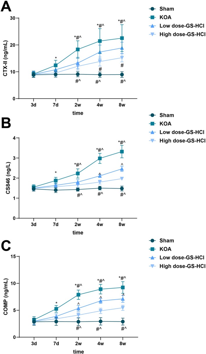
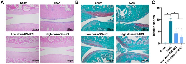
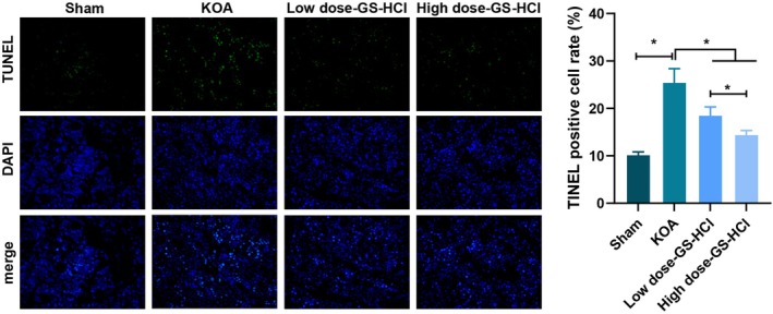
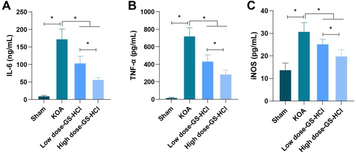
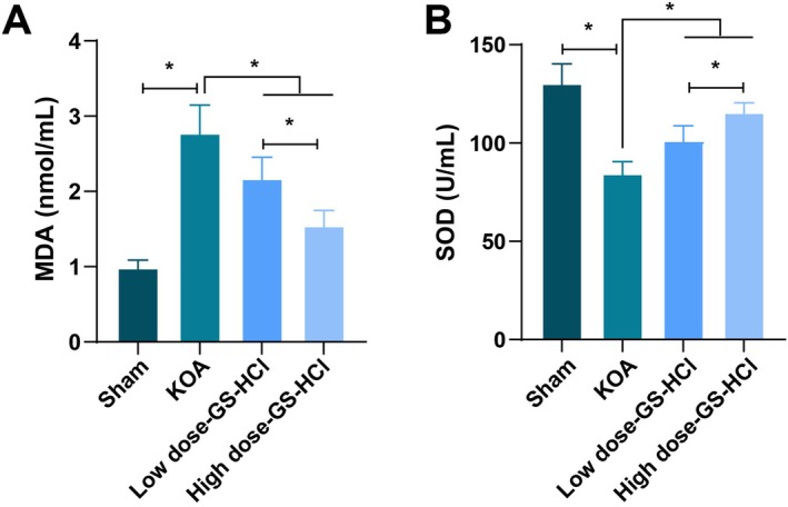
This study analysed the effects of different doses of glucosamine hydrochloride (GS-HCl) on cartilage tissue and the levels of joint injury markers in knee osteoarthritis (KOA). The Sham group, KOA group, low-dose GS-HCl group and high-dose GS-HCl group were established, with six mice in each group. The levels of joint injury markers (COMP, CS846 and CTX-II), inflammatory cytokines (IL-6, TNF-α and iNOS), oxidative stress indicators (MDA and SOD) and matrix remodelling proteins (MMP-3 and TIMP-1) were analysed. The degeneration of knee femoral condyles, histopathological changes and tissue apoptosis rate of the articular cartilage was also assessed. Mice in the KOA group displayed elevated COMP, CS846, CTX-II, IL-6, TNF-α, iNOS and MDA contents, reduced SOD activity, an irregular articular cartilage surface, a serious cartilage defect, a disordered articular cartilage surface in the defect, disappeared cartilage cells, obvious synovial cell proliferation and visible inflammatory cell infiltration. In the tissue, apoptosis rate and MMP-3 and TIMP-1 protein expression increased. Different doses of GS-HCl treatment could reduce COMP, CS846, CTX-II, IL-6, TNF-α, iNOS and MDA contents, apoptosis rate and MMP-3 and TIMP-1 protein expression, increase SOD activity and improve histopathological conditions in KOA mice. The improvement effects in each indicator in the high dose-GS-HCl group were more significant than those in the low dose-GS-HCl group. The intragastric administration of the GS-HCl group partially prevents the degeneration of articular cartilage in KOA mice. The mechanism may be to reduce inflammatory factors and oxidative stress indicator expression and matrix degradation, thereby delaying osteoarthritis progression.
本研究分析了不同剂量盐酸氨基葡萄糖(GS-HCl)对膝关节骨关节炎(KOA)软骨组织及关节损伤标志物水平的影响。建立了假手术组、KOA组、低剂量GS-HCl组和高剂量GS-HCl组,每组6只小鼠。分析了关节损伤标志物(COMP、CS846和CTX-II)、炎性细胞因子(IL-6、TNF-α和iNOS)、氧化应激指标(MDA和SOD)以及基质重塑蛋白(MMP-3和TIMP-1)的水平。还评估了膝关节股骨髁的退变、组织病理学变化以及关节软骨的组织凋亡率。KOA组小鼠的COMP、CS846、CTX-II、IL-6、TNF-α、iNOS和MDA含量升高,SOD活性降低,关节软骨表面不规则,软骨缺损严重,缺损处关节软骨表面紊乱,软骨细胞消失,滑膜细胞明显增殖,炎性细胞浸润可见。在组织中,凋亡率以及MMP-3和TIMP-1蛋白表达增加。不同剂量的GS-HCl治疗可降低KOA小鼠的COMP、CS846、CTX-II、IL-6、TNF-α、iNOS和MDA含量、凋亡率以及MMP-3和TIMP-1蛋白表达,增加SOD活性并改善组织病理学状况。高剂量GS-HCl组各指标的改善效果比低剂量GS-HCl组更显著。GS-HCl组灌胃给药可部分预防KOA小鼠关节软骨的退变。其机制可能是减少炎性因子和氧化应激指标的表达以及基质降解,从而延缓骨关节炎的进展。