Li Mingkai, Zhang Zhi, Chen Zebin, Chen Xi, Liu Huaqing, Xiao Yuanqiang, Chen Haimei, Zong Xiaodan, Chen Jingbiao, Chen Jianning, Wang Xinying, Xiao Xuehong, Yang Zhiwei, Han Lanqing, Wang Jin, Wu Bin
From the Department of Gastroenterology, The Third Affiliated Hospital of Sun Yat-sen University, No. 600 Tianhe Rd, Guangzhou 510000, China.
Tourism and Historical Culture College, Zhaoqing University, Zhaoqing, China.
Radiol Imaging Cancer. 2025 May;7(3):e240332. doi: 10.1148/rycan.240332.
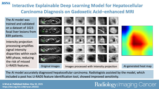
Purpose To develop an artificial intelligence (AI) model based on gadoxetic acid-enhanced MRI to assist radiologists in hepatocellular carcinoma (HCC) diagnosis. Materials and Methods This retrospective study included patients with focal liver lesions (FLLs) who underwent gadoxetic acid-enhanced MRI between January 2015 and December 2021. All hepatic malignancies were diagnosed pathologically, whereas benign lesions were confirmed with pathologic findings or imaging follow-up. Five manually labeled bounding boxes for each FLL obtained from precontrast T1-weighted, T2-weighted, arterial phase, portal venous phase, and hepatobiliary phase images were included. The lesion classifier component, used to distinguish HCC from non-HCC, was trained and externally tested. The feature classifier, based on a post hoc algorithm, inferred the presence of the Liver Imaging Reporting and Data System (LI-RADS) features by analyzing activation patterns of the pretrained lesion classifier. Two radiologists categorized FLLs in the external testing dataset according to LI-RADS criteria. Diagnostic performance of the AI model and the model's impact on reader accuracy were assessed. Results The study included 839 patients (mean age, 51 years ± 12 [SD]; 681 male) with 1023 FLLs (594 HCCs and 429 non-HCCs). The AI model yielded area under the receiver operating characteristic curves of 0.98 and 0.97 in the training set and external testing set, respectively. Compared with LI-RADS category 5, the AI model showed higher sensitivity (91.6% vs 74.8%; < .001) and similar specificity (90.7% vs 96.0%; = .22). The two readers identified more LI-RADS major features and more accurately classified category LR-5 lesions when assisted versus unassisted by AI, with higher sensitivities (reader 1, 85.7% vs 72.3%; < .001; reader 2, 89.1% vs 74.0%; < .001) and the same specificities (reader 1, 93.3% vs reader 2, 94.7%; > .99 for both). Conclusion The AI model accurately diagnosed HCC and improved the radiologists' diagnostic performance. Artificial Intelligence, Deep Learning, MRI, Hepatocellular Carcinoma © RSNA, 2025 See also commentary by Singh et al in this issue.
目的 开发一种基于钆塞酸增强磁共振成像(MRI)的人工智能(AI)模型,以协助放射科医生进行肝细胞癌(HCC)诊断。材料与方法 这项回顾性研究纳入了2015年1月至2021年12月期间接受钆塞酸增强MRI检查的肝脏局灶性病变(FLL)患者。所有肝脏恶性肿瘤均经病理诊断,良性病变通过病理结果或影像学随访得以证实。纳入从平扫T1加权、T2加权、动脉期、门静脉期和肝胆期图像中获取的每个FLL的五个手动标注的边界框。用于区分HCC与非HCC的病变分类器组件进行了训练和外部测试。基于事后算法的特征分类器通过分析预训练病变分类器的激活模式来推断肝脏影像报告和数据系统(LI-RADS)特征的存在。两名放射科医生根据LI-RADS标准对外部测试数据集中的FLL进行分类。评估了AI模型的诊断性能及其对阅片者准确性的影响。结果 该研究纳入了839例患者(平均年龄51岁±12[标准差];男性681例),共有1023个FLL(594个HCC和429个非HCC)。AI模型在训练集和外部测试集中的受试者操作特征曲线下面积分别为0.98和0.97。与LI-RADS 5类相比,AI模型显示出更高的敏感性(9