Zhang Haoyang, Wang Xiaocen, Zhang Xu, Ma Yeting, Bao Penglin, Yu Yanhui, Wang Yuru, Gong Pengtao, Zhang Nan, Lee Soon-Ok, Li Xin, Li Jianhua
State Key Laboratory for Diagnosis and Treatment of Severe Zoonotic Infectious Diseases, Key Laboratory for Zoonosis Research of the Ministry of Education, Institute of Zoonosis, and College of Veterinary Medicine, Jilin University, Changchun, China.
Second Affiliated Hospital, Jilin University, Changchun, China.
PLoS Negl Trop Dis. 2025 Jun 2;19(6):e0013164. doi: 10.1371/journal.pntd.0013164. eCollection 2025 Jun.
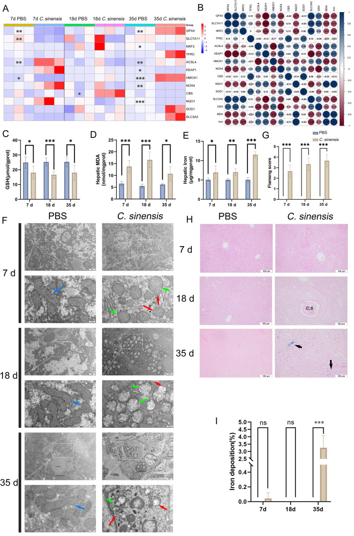
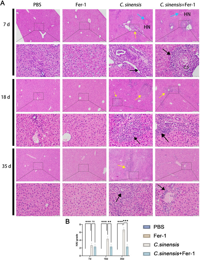
Clonorchis sinensis (C. sinensis) is a food-borne zoonotic parasite link to liver fibrosis and cholangiocarcinoma. Limited understanding of its mechanisms in causing liver fibrosis has impeded therapeutic advances for C. sinensis-induced liver lesions. Ferroptosis, a novel form of cell death involving iron overload and lipid peroxidation, exacerbates liver fibrosis; however, its role in C. sinensis infection remains unexplored. In this study, ferroptosis were detected in C. sinensis-infected C57BL/6 mice as well as in AML12 cells stimulated by C. sinensis excretory/secretory products (ESPs). 12 ferroptosis related genes were screened and we found glutathione peroxidase 4 (GPX4, 7 d), solute carrier family 7 member 11 (SLC7A11, 7 d) and nuclear factor erythroid 2 related factor 2 (Nrf2, 35 d) was significantly decreased in mice. Western blot results confirmed C. sinensis and ESPs down-regulated the expression of GPX4, SLC7A11 and Nrf2. GSH depletion, malondialdehyde (MDA) accumulation, mitochondrial structure damage, and iron overload were found in C. sinensis-infected mice and ESPs-stimulated AML12 cells, suggesting that ferroptosis occurred in vivo and in vitro. Treatment with ferroptosis inhibitor Fer-1 in C. sinensis-infected mice alleviated ferroptosis, reduced the productions of IFN-γ, TNF-α, IL-12 and IL-6, and downregulated transforming growth factor (TGF)-β/Smad pathway activation. In AML12 cells, Fer - 1 pretreatment reduced ESPs - induced ferroptosis and IL-6, TNF-α production. Fer - 1 also alleviated liver lesions, reduced parasite load (65%), α-SMA expression and collagen fiber deposition in infected mice. In conclusion, C. sinensis could cause ferroptosis, which promoted the secretions of IL-6 and TNF-α as well as the activation of TGF-β/Smad pathway, leading to exacerbated liver fibrosis.
华支睾吸虫是一种食源性人畜共患寄生虫,与肝纤维化和胆管癌有关。对其导致肝纤维化机制的了解有限,阻碍了华支睾吸虫所致肝脏病变治疗方面的进展。铁死亡是一种涉及铁过载和脂质过氧化的新型细胞死亡形式,会加剧肝纤维化;然而,其在华支睾吸虫感染中的作用仍未得到探索。在本研究中,在华支睾吸虫感染的C57BL/6小鼠以及受华支睾吸虫排泄/分泌产物(ESPs)刺激的AML12细胞中检测到了铁死亡。筛选出12个与铁死亡相关的基因,我们发现小鼠体内谷胱甘肽过氧化物酶4(GPX4,7天)、溶质载体家族7成员11(SLC7A11,7天)和核因子红细胞2相关因子2(Nrf2,35天)显著降低。蛋白质印迹结果证实华支睾吸虫和ESPs下调了GPX4、SLC7A11和Nrf2的表达。在华支睾吸虫感染的小鼠和ESPs刺激的AML12细胞中发现了谷胱甘肽(GSH)耗竭、丙二醛(MDA)积累、线粒体结构损伤和铁过载,表明体内和体外均发生了铁死亡。用铁死亡抑制剂Fer-1治疗华支睾吸虫感染的小鼠可减轻铁死亡,减少干扰素-γ、肿瘤坏死因子-α、白细胞介素-12和白细胞介素-6的产生,并下调转化生长因子(TGF)-β/Smad通路的激活。在AML12细胞中,Fer-1预处理减少了ESPs诱导的铁死亡以及白细胞介素-6、肿瘤坏死因子-α的产生。Fer-1还减轻了肝脏病变,降低了感染小鼠的寄生虫负荷(65%)、α-平滑肌肌动蛋白(α-SMA)表达和胶原纤维沉积。总之,华支睾吸虫可导致铁死亡,促进白细胞介素-6和肿瘤坏死因子-α的分泌以及TGF-β/Smad通路的激活,导致肝纤维化加剧。