Dong Fan, Lai Shanglei, Qiu Jiannan, Dou Xiaobing
School of Public Health, Zhejiang Chinese Medical University, Hangzhou 310053, China.
Medical Research Center, Shaoxing People's Hospital, Shaoxing 312000, Zhejiang Province, China.
Zhejiang Da Xue Xue Bao Yi Xue Ban. 2025 May 25;54(3):307-317. doi: 10.3724/zdxbyxb-2024-0381.
To explore the protective effect of vitexin-4 ″--glucoside (VOG) against acetaminophen-induced acute liver injury in mice and its underlying mechanism.
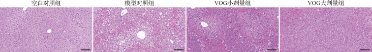
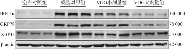
C57BL/6 mice were randomly divided into 4 groups: normal control group, model control group, low-dose group of VOG (30 mg/kg), and high-dose group of VOG (60 mg/kg). Acute liver injury was induced by intraperitoneal injection of acetaminophen (500 mg/kg). VOG was administrated by gavage 2 h before acetaminophen treatment in VOG groups. The protective effect of VOG against acute liver injury was evaluated by detecting alanine transaminase (ALT), aspartate transaminase (AST) levels and hematoxylin and eosin staining. The malondialdehyde (MDA) content, superoxide dismutase (SOD) and catalase (CAT) activity in liver were detected to evaluate the hepatic oxidative stress. The expression levels of tumor necrosis factor (TNF)-α, -, and - in liver were detected by quantitative reverse transcription polymerase chain reaction (qRT-PCR). The expression levels of phosphorylated c-jun -terminal kinase (JNK)/JNK, phosphorylated p38/p38, inositol-requiring enzyme 1 alpha (IRE-1α), X-box binding protein 1s (XBP1s), and glucose-regulated protein 78 (GRP78) in liver were detected by Western blotting. An endoplasmic reticulum stress model was established in AML-12 cells using tunicamycin. Cell viability was assessed using the CCK-8 assay, and the degree of cell damage was detected by lactate dehydrogenase (LDH) assay. The gene expression levels of -, , and in the cells were detected using qRT-PCR.
In the animal experiments, compared with the model control group, VOG significantly improved plasma ALT and AST levels, liver MDA content, as well as SOD and CAT activities. VOG also reduced the expression levels of -, -, and - in the liver, and improved protein phosphorylation levels of JNK and p38, as well as the protein expression levels of IRE-1α, XBP1s, and GRP78. In cell experiments, VOG pretreatment enhanced cell viability, reduced LDH release and decreased the mRNA expression of -, , and .
VOG can suppress inflammation and oxidative stress, and alleviate acetaminophen-induced acute liver injury in mice by suppressing endoplasmic reticulum stress and modulating the MAPK signaling pathway.
探讨牡荆素-4″-O-葡萄糖苷(VOG)对乙酰氨基酚诱导的小鼠急性肝损伤的保护作用及其潜在机制。
将C57BL/6小鼠随机分为4组:正常对照组、模型对照组、VOG低剂量组(30 mg/kg)和VOG高剂量组(60 mg/kg)。通过腹腔注射乙酰氨基酚(500 mg/kg)诱导急性肝损伤。在VOG组中,于乙酰氨基酚处理前2 h通过灌胃给予VOG。通过检测丙氨酸转氨酶(ALT)、天冬氨酸转氨酶(AST)水平及苏木精-伊红染色评估VOG对急性肝损伤的保护作用。检测肝脏中丙二醛(MDA)含量、超氧化物歧化酶(SOD)和过氧化氢酶(CAT)活性以评估肝脏氧化应激。通过定量逆转录聚合酶链反应(qRT-PCR)检测肝脏中肿瘤坏死因子(TNF)-α等的表达水平。通过蛋白质印迹法检测肝脏中磷酸化的c-jun氨基末端激酶(JNK)/JNK、磷酸化的p38/p38、肌醇需要酶1α(IRE-1α)、X盒结合蛋白1s(XBP1s)和葡萄糖调节蛋白78(GRP78)的表达水平。使用衣霉素在AML-12细胞中建立内质网应激模型。使用CCK-8法评估细胞活力,并通过乳酸脱氢酶(LDH)测定法检测细胞损伤程度。使用qRT-PCR检测细胞中相关基因的表达水平。
在动物实验中,与模型对照组相比,VOG显著改善了血浆ALT和AST水平、肝脏MDA含量以及SOD和CAT活性。VOG还降低了肝脏中TNF-α等的表达水平,并改善了JNK和p38的蛋白磷酸化水平以及IRE-1α、XBP1s和GRP78的蛋白表达水平。在细胞实验中,VOG预处理增强了细胞活力,降低了LDH释放,并降低了相关基因的mRNA表达。
VOG可通过抑制内质网应激和调节丝裂原活化蛋白激酶(MAPK)信号通路来抑制炎症和氧化应激,并减轻乙酰氨基酚诱导的小鼠急性肝损伤。