van den Berg Ingeborg, Zachiu Cornel, de Groot-van Breugel Eline N, Willigenburg Thomas, Bol Gijsbert H, Lagendijk Jan J W, Raaymakers Bas W, van Melick Harm H E, van den Berg Cornelis A T, van der Voort van Zyp Jochem R N, de Boer Johannes C J
Department of Radiation Oncology, Division of Imaging & Oncology, University Medical Center Utrecht, Utrecht, The Netherlands.
Department of Urology, St. Antonius Hospital, Utrecht/Nieuwegein, The Netherlands.
Phys Imaging Radiat Oncol. 2025 May 10;34:100776. doi: 10.1016/j.phro.2025.100776. eCollection 2025 Apr.
A sub-fractionation workflow enables a substantial reduction in planning target volume (PTV) margin in prostate cancer (PCa) patients by reducing systematic motion during magnetic resonance (MR)-guided radiotherapy. This study assessed geometric and reconstructed dose outcomes in patients treated with a tight-margin sub-fractionation workflow on a combined linear accelerator with a 1.5 T MRI scanner (MR-Linac).
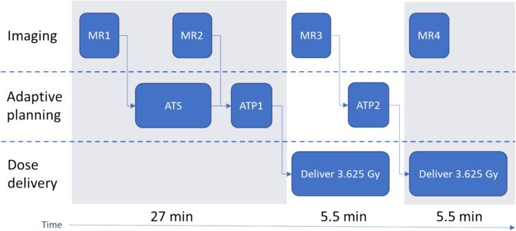
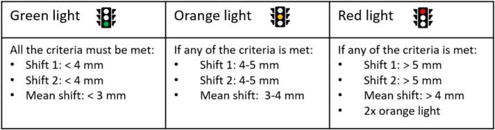
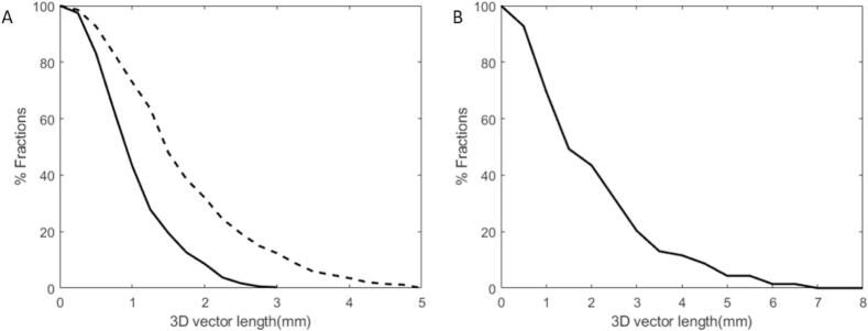
We evaluated the sub-fractionation workflow with tight margins (2-3 mm) on 128 PCa patients who completed treatment with 5 × 7.25 Gy (36.25 Gy total dose). A traffic light protocol was applied based on residual motions to detect patients with unexpectedly large motions. When 'red' traffic light criteria were met, plans with larger margins (5 mm isotropic) were adopted for subsequent fractions. Intra- and inter-fraction dose accumulation was performed via an in-house developed deformable image registration algorithm.
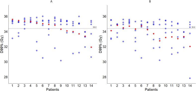
A total of 89 % (114/128) of patients completed treatment with the initial tight margins. The mean 3D intrafraction shifts were 1.0 mm (SD: 0.6 mm) in the group with the tight margins and 1.9 mm (SD: 1.5 mm) in the patient group who switched to large margins. The median accumulated D99% was 34.9 Gy (interquartile range: 34.0-35.3 Gy) for patients with prostate shifts who switched to larger margins. In 57 % (8/14) of these patients, the accumulated D99% was above the threshold of 34.4 Gy.
Tight margins of 2-3 mm can be safely applied for at least 95 % (122/128) of the PCa patients undergoing a sub-fractionation workflow on a 1.5 T MR-linac.
通过减少磁共振(MR)引导放疗期间的系统运动,子分割工作流程能够大幅降低前列腺癌(PCa)患者的计划靶区(PTV)边界。本研究评估了在配备1.5T MRI扫描仪的组合直线加速器(MR-Linac)上采用窄边界子分割工作流程治疗的患者的几何和重建剂量结果。
我们对128例完成5×7.25Gy(总剂量36.25Gy)治疗的PCa患者评估了窄边界(2-3mm)的子分割工作流程。基于残余运动应用交通灯协议来检测运动意外较大的患者。当满足“红色”交通灯标准时,后续分次采用边界更大(各向同性5mm)的计划。通过内部开发的可变形图像配准算法进行分次内和分次间剂量累积。
共有89%(114/128)的患者以初始窄边界完成治疗。窄边界组的平均三维分次内位移为1.0mm(标准差:0.6mm),而转为宽边界的患者组为1.9mm(标准差:1.5mm)。转为宽边界的前列腺移位患者的中位累积D99%为34.9Gy(四分位间距:34.0-35.3Gy)。在这些患者中,57%(8/14)的累积D99%高于34.4Gy的阈值。
对于在1.5T MR-Linac上接受子分割工作流程的PCa患者,至少95%(122/128)可以安全地应用2-3mm的窄边界。