Sitzman Thomas J, Chee-Williams Jessica L, Snodgrass Taylor D, Gilbert Imani R, Tollefson Travis T, Singh Davinder J, Perry Jamie L
From the Division of Plastic Surgery, Center for Cleft and Craniofacial Care, Phoenix Children's Hospital, Phoenix, AZ.
Division of Plastic Surgery, Mayo Clinic Arizona, Scottsdale, AZ.
Plast Reconstr Surg Glob Open. 2025 Jun 16;13(6):e6797. doi: 10.1097/GOX.0000000000006797. eCollection 2025 Jun.
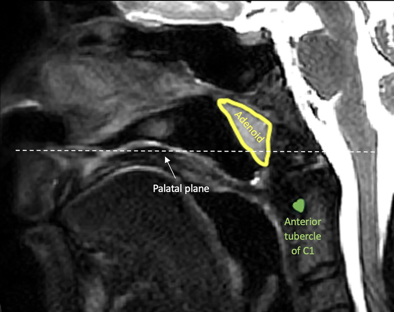
Positioning the pharyngeal flap base high along the posterior pharyngeal wall is essential for optimizing speech outcomes. Objective data on where to place the flap base are lacking. Further, adenoid tissue can restrict cephalad positioning of the flap. This study aimed to improve the design of the pharyngeal flap by measuring the distance from the first cervical vertebrae (C1) to the palatal plane, and the adenoid depth in children undergoing evaluation for velopharyngeal insufficiency.
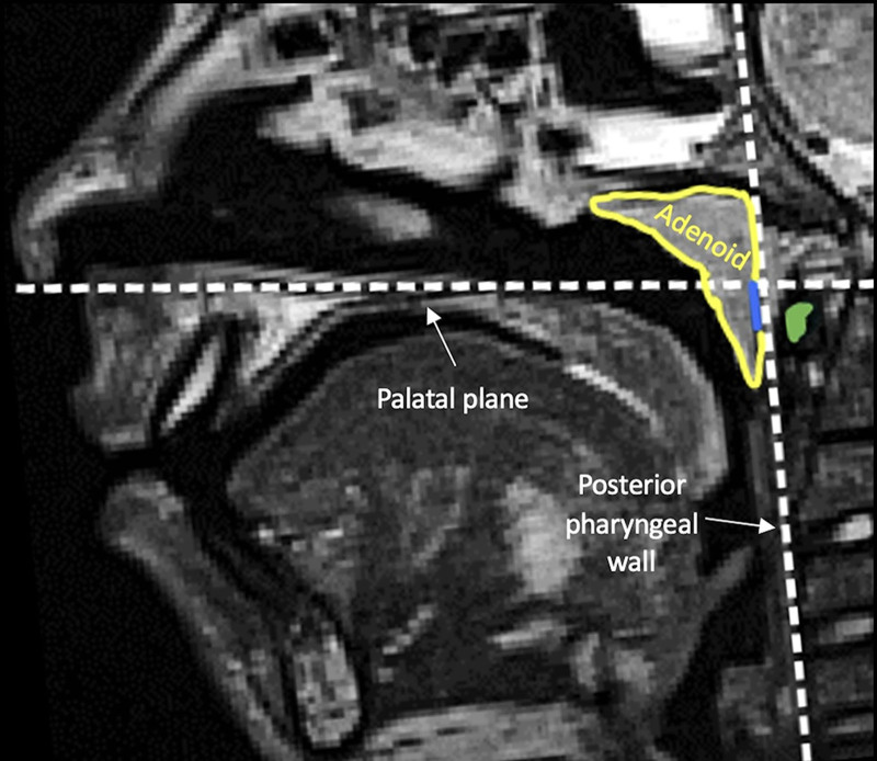
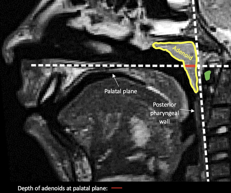
This retrospective cross-sectional study analyzed magnetic resonance imaging (MRI) scans of the velopharynx and measured the distance between C1 and the palatal plane, and the adenoid depth at the level of the palatal plane in millimeters.
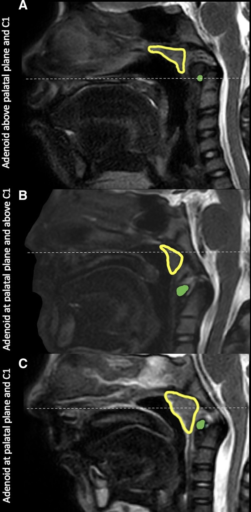
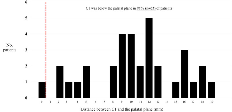
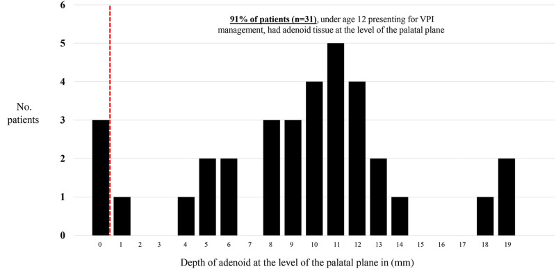
Thirty-four patients met the inclusion criteria. The mean age at the time of MRI was 7.4 years (range: 3.9-11.9 y). The anterior tubercle of C1 was below the palatal plane in 97% (n = 33) of patients. On average, this landmark was 10.5 mm (SD = 5.0) below the palatal plane. Adenoid tissue was present at the level of the palatal plane in 91% (n = 31) of patients.
Positioning the pharyngeal flap base at C1 is too low to aid with velopharyngeal closure. Further, adenoid tissue is frequently present at the level of velopharyngeal closure, limiting superior positioning of the pharyngeal flap base. When this occurs, surgeons should consider adenoidectomy before pharyngeal flap surgery. Preoperative MRI may be beneficial for planning pharyngeal flap positioning relative to C1 and assessing adenoid tissue at the palatal plane.
将咽瓣基底部沿咽后壁高位定位对于优化语音效果至关重要。目前缺乏关于咽瓣基底部放置位置的客观数据。此外,腺样体组织会限制咽瓣向上的定位。本研究旨在通过测量接受腭咽闭合不全评估的儿童中第一颈椎(C1)至腭平面的距离以及腺样体深度,来改进咽瓣的设计。
这项回顾性横断面研究分析了腭咽部的磁共振成像(MRI)扫描图像,并测量了C1与腭平面之间的距离以及腭平面水平处腺样体的深度(以毫米为单位)。
34例患者符合纳入标准。MRI检查时的平均年龄为7.4岁(范围:3.9 - 11.9岁)。97%(n = 33)的患者C1的前结节位于腭平面下方。平均而言,该标志点位于腭平面下方10.5毫米(标准差 = 5.0)。91%(n = 31)的患者在腭平面水平存在腺样体组织。
将咽瓣基底部定位在C1处过低,无法辅助腭咽闭合。此外,在腭咽闭合水平经常存在腺样体组织,限制了咽瓣基底部向上定位。当出现这种情况时,外科医生在进行咽瓣手术前应考虑腺样体切除术。术前MRI对于规划咽瓣相对于C1的定位以及评估腭平面处的腺样体组织可能有益。