Freni Josè, Centofanti Antonio, Nicita Fabiana, Labellarte Davide, Vermiglio Giovanna, Anastasi Michele Runci
Department of Biomedical, Dental Sciences and Morphofunctional Imaging, University of Messina, 98122 Messina, Italy.
Department of Maxillo-Facial Surgery, University of Sapienza, 00161 Roma, Italy.
J Funct Morphol Kinesiol. 2025 Mar 27;10(2):107. doi: 10.3390/jfmk10020107.
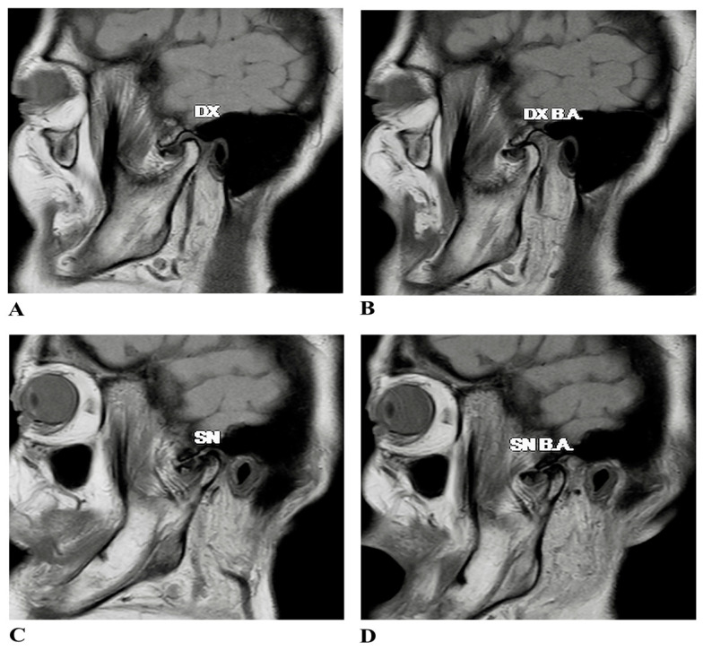
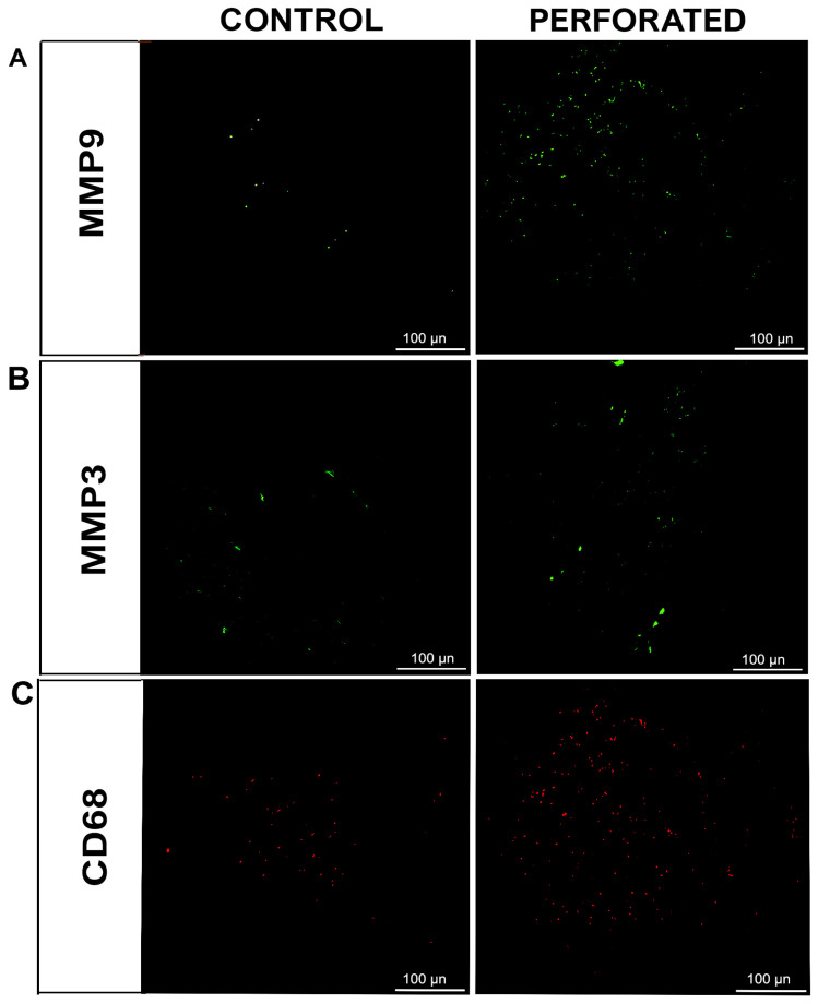
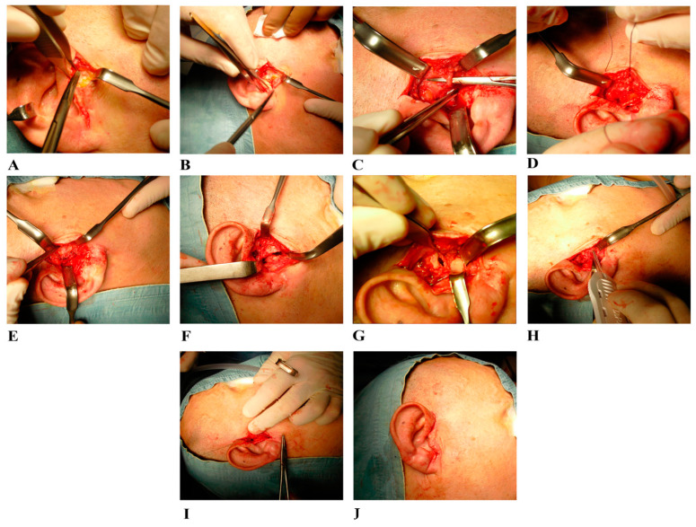
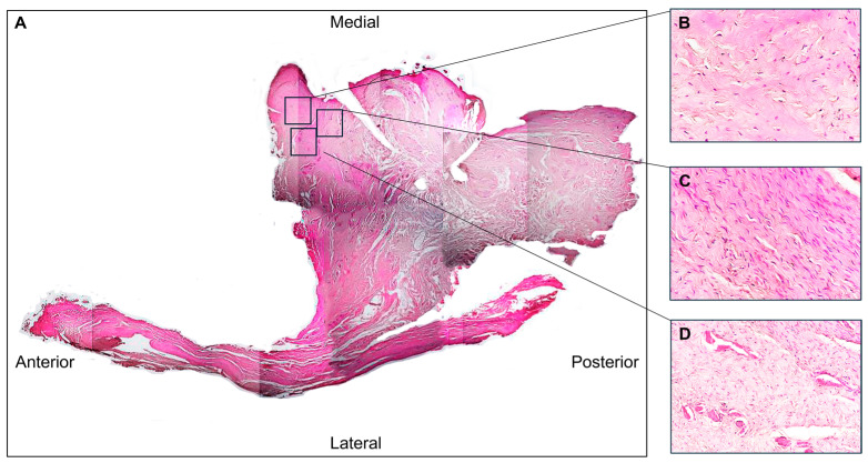
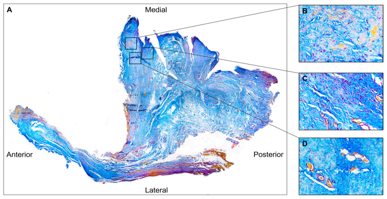
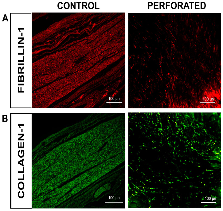
: Anterior disc displacement without reduction (ADDwoR) is a temporomandibular joint (TMJ) disorder characterized by progressive dysfunction and potential complications. Persistent displacement leads to abnormal mechanical stress, predisposing the TMJ disc to structural degeneration, including perforation. This case report aimed to examine the histological and immunofluorescence characteristics of perforated disc tissue to elucidate the mechanisms contributing to its pathology. : A 50-year-old patient with bilateral ADDwoR and disc perforation underwent functional arthroplasty. Tissue samples from the perforated disc were histologically analyzed using hematoxylin-eosin and Azan Mallory staining. Immunofluorescence was performed to assess the expression of collagen type I, fibrillin-1, matrix metalloproteinases (MMPs)-3 and -9, and cluster of differentiation 68 (CD68). : Histological analysis revealed disorganized collagen fibres and fibro-chondrocyte cell predominance in the perilesional zone, accompanied by vascular proliferation. Adjacent tissue to perforation exhibited normal fibrous organization. Immunofluorescence showed reduced collagen type I and fibrillin-1 patterns in the perilesional area, indicating an alteration in the fibrillar component of the extracellular matrix (ECM). Increased expression of MMP-3 and MMP-9, as well as elevated numbers of CD68-positive macrophages, suggested active ECM degradation and inflammation localized to the perforation site. : This case report underscores the critical role of biomechanical stress and inflammation in disc perforation. Decreased ECM integrity, driven by altered collagen and fibrillin composition, as well as heightened MMP activity, compromises the disc's capacity to absorb and distribute mechanical loads. These findings advance our understanding of TMJ pathophysiology, emphasizing the need for therapeutic approaches that target both biomechanical stabilization and inflammation.
不可复性盘前移位(ADDwoR)是一种颞下颌关节(TMJ)疾病,其特征为进行性功能障碍和潜在并发症。持续性移位会导致异常机械应力,使TMJ盘易于发生结构退变,包括穿孔。本病例报告旨在研究穿孔盘组织的组织学和免疫荧光特征,以阐明其病理形成机制。
一名患有双侧ADDwoR和盘穿孔的50岁患者接受了功能性关节成形术。对穿孔盘的组织样本进行苏木精-伊红和阿赞-马洛里染色的组织学分析。进行免疫荧光检测以评估I型胶原、原纤蛋白-1、基质金属蛋白酶(MMP)-3和-9以及分化簇68(CD68)的表达。
组织学分析显示,病变周围区域胶原纤维排列紊乱,以纤维软骨细胞为主,并伴有血管增生。穿孔相邻组织表现出正常的纤维组织。免疫荧光显示病变周围区域I型胶原和原纤蛋白-1的模式减少,表明细胞外基质(ECM)的纤维成分发生改变。MMP-3和MMP-9表达增加,以及CD68阳性巨噬细胞数量增多,提示ECM的主动降解和炎症局限于穿孔部位。
本病例报告强调了生物力学应力和炎症在盘穿孔中的关键作用。胶原和原纤蛋白组成改变导致ECM完整性降低,以及MMP活性增强,损害了盘吸收和分布机械负荷的能力。这些发现增进了我们对TMJ病理生理学的理解,强调了针对生物力学稳定和炎症的治疗方法的必要性。