Tan Liangzhang, Liu Shoukuan, Linghu Wenrui, Ke Yifeng, Li Yongtao, Pazo Emmanuel Eric, Ren Xinjun
Tianjin Key Laboratory of Retinal Functions and Diseases, Tianjin Branch of National Clinical Research Center for Ocular Disease, Eye Institute and School of Optometry, Tianjin Medical University Eye Hospital, Tianjin, China.
BMC Ophthalmol. 2025 Jul 1;25(1):374. doi: 10.1186/s12886-025-04186-6.
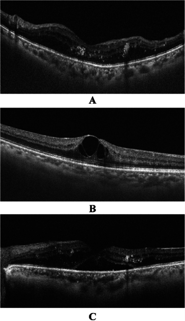
This study aimed to observe the morphological changes and visual outcomes in diabetic macular edema (DME) subtypes based on optical coherence tomography classification following intravitreal injection.
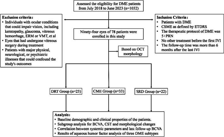
Ninety-four eyes from seventy-eight patients were enrolled between July 1, 2018, and June 30, 2023. Best-corrected visual acuity (BCVA), central subfield thickness, and morphological changes were examined at each follow-up visit. Aqueous humor samples were obtained before intravitreal injection of anti-vascular endothelial growth factor or cataract surgery. The inflammatory cytokine profile in the aqueous humor of each DME subtype was analyzed to explore potential contributions.
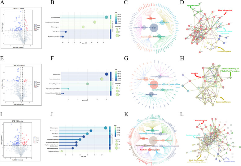
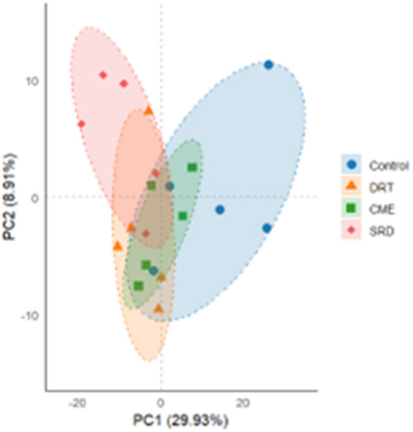
The BCVA in the diffuse retinal thickening group was significantly improved at the last follow-up (p = 0.006), but no significant change in central subfield thickness was observed. Conversely, the BCVA in the cystoid macular edema and serous retinal detachment groups did not significantly improve, although a significant reduction in central subfield thickness was observed in both groups at the final follow-up (both p < 0.05). Principal component analysis of proteomic data revealed clear segregation between samples from patients with different DME subtypes and those from the control group. 59 proteins were found to be significantly regulated in the diffuse retinal thickening group compared to those in the control group. Of these, 10 proteins were upregulated, whereas 49 were downregulated. In the cystoid macular edema group, 30 proteins were significantly regulated, with 8 proteins upregulated and 22 downregulated. In the serous retinal detachment group, 87 proteins showed significant changes: 24 exhibited increased expression and 63 decreased expression.
The diffuse retinal thickening subtype showed a better response and cystoid macular edema was the most common morphological subtype of DME after intravitreal injection treatment in real-world clinical observations. The variations in protein expression of aqueous humor suggested distinct pathological mechanisms for each DME subtype, laying the groundwork for more targeted treatment strategies.
本研究旨在观察玻璃体内注射后基于光学相干断层扫描分类的糖尿病性黄斑水肿(DME)亚型的形态学变化和视觉结果。
2018年7月1日至2023年6月30日期间纳入了78例患者的94只眼。每次随访时检查最佳矫正视力(BCVA)、中心子野厚度和形态学变化。在玻璃体内注射抗血管内皮生长因子或白内障手术前采集房水样本。分析各DME亚型房水中的炎性细胞因子谱,以探索潜在作用。
弥漫性视网膜增厚组在末次随访时BCVA显著改善(p = 0.006),但中心子野厚度未观察到显著变化。相反,囊样黄斑水肿和浆液性视网膜脱离组的BCVA未显著改善,尽管两组在末次随访时中心子野厚度均显著降低(均p < 0.05)。蛋白质组学数据的主成分分析显示,不同DME亚型患者的样本与对照组样本之间存在明显分离。与对照组相比,弥漫性视网膜增厚组有59种蛋白质被显著调节。其中,10种蛋白质上调,49种蛋白质下调。在囊样黄斑水肿组中,30种蛋白质被显著调节,8种蛋白质上调,22种蛋白质下调。在浆液性视网膜脱离组中,87种蛋白质有显著变化:24种表达增加,63种表达减少。
在真实世界临床观察中,玻璃体内注射治疗后,弥漫性视网膜增厚亚型显示出更好的反应,囊样黄斑水肿是DME最常见的形态学亚型。房水蛋白质表达的差异提示各DME亚型有不同的病理机制,为更具针对性的治疗策略奠定了基础。