• 文献检索
  • 文档翻译
  • 深度研究
  • 学术资讯
  • Suppr Zotero 插件Zotero 插件
  • 邀请有礼
  • 套餐&价格
  • 历史记录
应用&插件
Suppr Zotero 插件Zotero 插件浏览器插件Mac 客户端Windows 客户端微信小程序
定价
高级版会员购买积分包购买API积分包
服务
文献检索文档翻译深度研究API 文档MCP 服务
关于我们
关于 Suppr公司介绍联系我们用户协议隐私条款
关注我们

Suppr 超能文献

核心技术专利:CN118964589B侵权必究
粤ICP备2023148730 号-1Suppr @ 2026

文献检索

告别复杂PubMed语法,用中文像聊天一样搜索,搜遍4000万医学文献。AI智能推荐,让科研检索更轻松。

立即免费搜索

文件翻译

保留排版,准确专业,支持PDF/Word/PPT等文件格式,支持 12+语言互译。

免费翻译文档

深度研究

AI帮你快速写综述,25分钟生成高质量综述,智能提取关键信息,辅助科研写作。

立即免费体验

在无临床视网膜病变证据的糖尿病患者中,视网膜中周部毛细血管无灌注区扩大。

Retinal Mid-Peripheral Capillary Free Zones Are Enlarged in Diabetic Patients With No Evidence of Clinical Retinopathy.

作者信息

Arthur Edmund, Ravichandran Swetha, Rothstein Andrew, Brown Keisha, Jonnadula Ganesh B, Murchison Charles F, Grant Maria B

机构信息

School of Optometry, University of Alabama at Birmingham, Birmingham, AL, USA.

Department of Neurology, Heersink School of Medicine, University of Alabama at Birmingham, Birmingham, AL, USA.

出版信息

Transl Vis Sci Technol. 2025 Jul 1;14(7):10. doi: 10.1167/tvst.14.7.10.

DOI:10.1167/tvst.14.7.10
PMID:40673733
原文链接:https://pmc.ncbi.nlm.nih.gov/articles/PMC12282639/
Abstract

PURPOSE

To compare the width of the mid-peripheral capillary free zones (CFZs; periarteriole and perivenule) between diabetics with no diabetic retinopathy (DR) versus controls.

METHODS

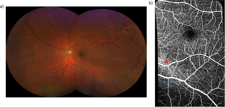
The 20° × 20° optical coherence tomography angiography images of paired arterioles, venules, and their adjacent capillaries within the macular and inferomacular regions of the superficial vascular plexus were obtained from 46 eyes of 28 diabetics with no DR (mean age, 59 years; range, 40-71 years) and 46 eyes of 31 controls (mean age, 59 years; range, 46-78 years). There was no significant difference in age between groups (P = 0.77). The macular and inferomacular images were montaged to generate a wider field of view, followed by the application of a vesselness filter and Otsu thresholding. The mid-peripheral CFZ width was calculated using previously established MATLAB algorithms. Generalized linear mixed models were used to compare the mid-peripheral CFZs between groups, accounting for correlation between eyes.

RESULTS

The periarteriole CFZ width was greater in diabetics with no DR (73.3 ± 6.49 µm) compared to controls (67.3 ± 7.08 µm; P < 0.001, Cohen's d = 0.88). Similarly, the perivenule CFZ width was larger in diabetics with no DR (60.8 ± 6.40 µm) compared to controls (54.8 ± 4.58 µm; P < 0.001, Cohens' d = 1.08).

CONCLUSIONS

Our results demonstrate larger mid-peripheral CFZ width in diabetics with no DR. The mid-peripheral CFZs show promise as a potential novel retinal vascular biomarker for early DR detection.

TRANSLATIONAL RELEVANCE

Our study shows the potential clinical utility of the mid-peripheral CFZs for early DR detection.

摘要

目的

比较无糖尿病视网膜病变(DR)的糖尿病患者与对照组中周毛细血管无灌注区(CFZ;小动脉周围和小静脉周围)的宽度。

方法

从28例无DR的糖尿病患者(平均年龄59岁;范围40 - 71岁)的46只眼中,以及31例对照组(平均年龄59岁;范围46 - 78岁)的46只眼中,获取浅表血管丛黄斑和黄斑下区域内成对小动脉、小静脉及其相邻毛细血管的20°×20°光学相干断层扫描血管造影图像。两组间年龄无显著差异(P = 0.77)。将黄斑和黄斑下图像拼接以生成更宽的视野,随后应用血管增强滤波器和大津阈值法。使用先前建立的MATLAB算法计算中周CFZ宽度。采用广义线性混合模型比较两组间的中周CFZ,同时考虑眼间的相关性。

结果

无DR的糖尿病患者小动脉周围CFZ宽度(73.3±6.49μm)大于对照组(67.3±7.08μm;P < 0.001;Cohen's d = 0.88)。同样,无DR的糖尿病患者小静脉周围CFZ宽度(60.8±6.40μm)大于对照组(54.8±4.58μm;P < 0.001;Cohens' d = 1.08)。

结论

我们的结果表明,无DR的糖尿病患者中周CFZ宽度更大。中周CFZ有望成为早期DR检测的潜在新型视网膜血管生物标志物。

转化相关性

我们的研究表明中周CFZ在早期DR检测中的潜在临床应用价值。

https://cdn.ncbi.nlm.nih.gov/pmc/blobs/a159/12282639/2fadaa0917bc/tvst-14-7-10-f007.jpg
https://cdn.ncbi.nlm.nih.gov/pmc/blobs/a159/12282639/8d51c7355a53/tvst-14-7-10-f001.jpg
https://cdn.ncbi.nlm.nih.gov/pmc/blobs/a159/12282639/a3370e35c3fc/tvst-14-7-10-f002.jpg
https://cdn.ncbi.nlm.nih.gov/pmc/blobs/a159/12282639/e07b8aaab4fa/tvst-14-7-10-f003.jpg
https://cdn.ncbi.nlm.nih.gov/pmc/blobs/a159/12282639/f89d36c85f69/tvst-14-7-10-f004.jpg
https://cdn.ncbi.nlm.nih.gov/pmc/blobs/a159/12282639/ad690ea0ab63/tvst-14-7-10-f005.jpg
https://cdn.ncbi.nlm.nih.gov/pmc/blobs/a159/12282639/8050f84f2fdf/tvst-14-7-10-f006.jpg
https://cdn.ncbi.nlm.nih.gov/pmc/blobs/a159/12282639/2fadaa0917bc/tvst-14-7-10-f007.jpg

相似文献

1
Retinal Mid-Peripheral Capillary Free Zones Are Enlarged in Diabetic Patients With No Evidence of Clinical Retinopathy.在无临床视网膜病变证据的糖尿病患者中,视网膜中周部毛细血管无灌注区扩大。
Transl Vis Sci Technol. 2025 Jul 1;14(7):10. doi: 10.1167/tvst.14.7.10.
2
Retinal mid-peripheral capillary free zones are enlarged in cognitively unimpaired older adults at high risk for Alzheimer's disease.认知正常的高龄阿尔茨海默病高危人群存在视网膜周边中部毛细血管无灌注区扩大。
Alzheimers Res Ther. 2023 Oct 12;15(1):172. doi: 10.1186/s13195-023-01312-8.
3
Optical coherence tomography (OCT) for detection of macular oedema in patients with diabetic retinopathy.光学相干断层扫描(OCT)用于检测糖尿病视网膜病变患者的黄斑水肿。
Cochrane Database Syst Rev. 2015 Jan 7;1(1):CD008081. doi: 10.1002/14651858.CD008081.pub3.
4
Optical coherence tomography (OCT) for detection of macular oedema in patients with diabetic retinopathy.光学相干断层扫描(OCT)用于检测糖尿病视网膜病变患者的黄斑水肿。
Cochrane Database Syst Rev. 2011 Jul 6(7):CD008081. doi: 10.1002/14651858.CD008081.pub2.
5
Impact of Anatomically Defined Retinal Zones on Ultrawide-Field Fluorescein Angiography Nonperfusion Measurements in Diabetic Retinopathy.解剖学定义的视网膜区域对糖尿病视网膜病变超广角荧光素血管造影无灌注测量的影响。
Ophthalmologica. 2025;248(3):194-201. doi: 10.1159/000545754. Epub 2025 Apr 15.
6
Clinical and imaging characteristics associated with foveal neovascularization in proliferative diabetic retinopathy.增殖性糖尿病视网膜病变中与黄斑中心凹新生血管相关的临床和影像学特征。
Graefes Arch Clin Exp Ophthalmol. 2025 Mar;263(3):679-687. doi: 10.1007/s00417-024-06660-1. Epub 2024 Nov 15.
7
Investigation of the impact of choroidal capillary flow area on macular ischemia in diabetic patients.脉络膜毛细血管血流面积对糖尿病患者黄斑缺血影响的研究。
Sci Rep. 2025 Jul 2;15(1):23615. doi: 10.1038/s41598-025-07130-2.
8
The Impact of Hypertension on Macular Perfusion in Patients With Referable Diabetic Retinopathy: An OCTA Analysis.高血压对可转诊糖尿病视网膜病变患者黄斑灌注的影响:一项光学相干断层扫描血管造影分析
Transl Vis Sci Technol. 2025 Jul 1;14(7):2. doi: 10.1167/tvst.14.7.2.
9
Anti-vascular endothelial growth factor for diabetic macular oedema: a network meta-analysis.抗血管内皮生长因子治疗糖尿病性黄斑水肿:一项网状Meta分析。
Cochrane Database Syst Rev. 2017 Jun 22;6(6):CD007419. doi: 10.1002/14651858.CD007419.pub5.
10
Artificial Intelligence Versus Rules-Based Approach for Segmenting NonPerfusion Area in a DRCR Retina Network Optical Coherence Tomography Angiography Dataset.在DRCR视网膜网络光学相干断层扫描血管造影数据集中,用于分割无灌注区的人工智能与基于规则的方法对比
Invest Ophthalmol Vis Sci. 2025 Mar 3;66(3):22. doi: 10.1167/iovs.66.3.22.

本文引用的文献

1
Quantifying Putative Retinal Gliosis in Preclinical Alzheimer's Disease.定量临床前阿尔茨海默病中的疑似视网膜神经胶质增生。
Invest Ophthalmol Vis Sci. 2024 May 1;65(5):5. doi: 10.1167/iovs.65.5.5.
2
Retinal mid-peripheral capillary free zones are enlarged in cognitively unimpaired older adults at high risk for Alzheimer's disease.认知正常的高龄阿尔茨海默病高危人群存在视网膜周边中部毛细血管无灌注区扩大。
Alzheimers Res Ther. 2023 Oct 12;15(1):172. doi: 10.1186/s13195-023-01312-8.
3
OCTA reveals remodeling of the peripheral capillary free zones in normal aging.
OCTA 显示正常衰老中周边毛细血管无灌注区的重构。
Sci Rep. 2021 Aug 2;11(1):15593. doi: 10.1038/s41598-021-95230-0.
4
Widefield Optical Coherence Tomography Angiography in Diabetic Retinopathy.宽视野光学相干断层扫描血管造影在糖尿病视网膜病变中的应用。
J Diabetes Res. 2020 Nov 24;2020:8855709. doi: 10.1155/2020/8855709. eCollection 2020.
5
Portable, non-invasive video imaging of retinal blood flow dynamics.便携式、非侵入性的视网膜血流动力学视频成像。
Sci Rep. 2020 Nov 19;10(1):20236. doi: 10.1038/s41598-020-76407-5.
6
Geometric Perfusion Deficits: A Novel OCT Angiography Biomarker for Diabetic Retinopathy Based on Oxygen Diffusion.几何性灌注缺损:基于氧扩散的糖尿病视网膜病变新型 OCT 血管造影生物标志物
Am J Ophthalmol. 2021 Feb;222:256-270. doi: 10.1016/j.ajo.2020.09.007. Epub 2020 Sep 9.
7
Optical Coherence Tomography Angiography Projection Artifact Removal: Impact on Capillary Density and Interaction with Diabetic Retinopathy Severity.光学相干断层扫描血管造影投影伪影去除:对毛细血管密度的影响及其与糖尿病视网膜病变严重程度的相互作用。
Transl Vis Sci Technol. 2020 Jun 5;9(7):10. doi: 10.1167/tvst.9.7.10. eCollection 2020 Jun.
8
Optical Coherence Tomography Angiography Avascular Area Association With 1-Year Treatment Requirement and Disease Progression in Diabetic Retinopathy.光学相干断层扫描血管造影无血管区与糖尿病视网膜病变 1 年治疗需求和疾病进展的关系。
Am J Ophthalmol. 2020 Sep;217:268-277. doi: 10.1016/j.ajo.2020.04.024. Epub 2020 Apr 29.
9
Distances From Capillaries to Arterioles or Venules Measured Using OCTA and AOSLO.使用 OCTA 和 AOSLO 测量毛细血管到小动脉或小静脉的距离。
Invest Ophthalmol Vis Sci. 2019 May 1;60(6):1833-1844. doi: 10.1167/iovs.18-25294.
10
Earliest Evidence of Preclinical Diabetic Retinopathy Revealed Using Optical Coherence Tomography Angiography Perfused Capillary Density.应用光相干断层扫描血管造影灌注毛细血管密度揭示临床前期糖尿病视网膜病变的最早证据。
Am J Ophthalmol. 2019 Jul;203:103-115. doi: 10.1016/j.ajo.2019.01.012. Epub 2019 Jan 26.