Whitmarsh Tristan
Institute of Astronomy, University of Cambridge, Madingley Rd, Cambridge CB3 0HA, United Kingdom.
JBMR Plus. 2025 Apr 28;9(9):ziaf075. doi: 10.1093/jbmrpl/ziaf075. eCollection 2025 Sep.
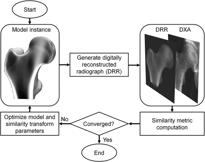
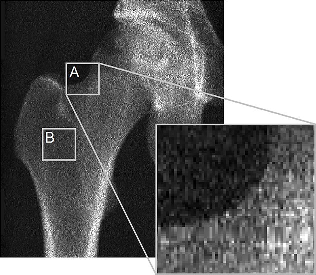
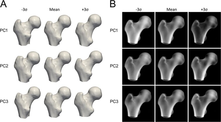
3D-DXA, as implemented in the software tool 3D-Shaper, is a software method that generates a 3D reconstruction of the proximal femur from a single 2D DXA image by registering a statistical model. Implementations of 3D-DXA aim to provide estimates of trabecular, cortical, and structural parameters, similar to those derived from quantitative computed tomography (QCT). As the inventor and developer of the software methods upon which 3D-DXA is built, I have been observing its adoption and widespread use with increasing concern. This article provides a critical evaluation of the methodological limitations inherent to 3D-DXA and discusses their implications for research and patient care. The primary issue is that the limited visibility of the cortex in a DXA image prevents 3D-DXA from accurately deriving cortical parameters. Instead, the software relies on predictions based on overall BMD rather than direct cortical measurements. This may lead to results that do not reflect actual cortical measurements. Additional concerns include the population bias due to the statistical model being derived from a specific demographic, and limited reconstruction accuracy by using single-view DXA images. These limitations have likely resulted in incorrect measurements and research outcomes, which have largely gone unrecognized due to the use of inappropriate performance assessment metrics and the absence of multiple comparison corrections in studies involving 3D-DXA. Despite these limitations, 3D-DXA has received regulatory approval in various countries, potentially compromising the accuracy of clinical diagnoses and treatment decisions. By highlighting these issues, this article aims to inform clinicians, researchers, and regulatory bodies about the significant limitations of 3D-DXA. It underscores the urgent need for a reevaluation of its use in research and clinical settings to prevent misinterpretation of results and to ensure patient safety.
软件工具3D-Shaper中所实现的三维双能X线吸收测定法(3D-DXA)是一种软件方法,通过配准统计模型从单个二维双能X线吸收测定(DXA)图像生成近端股骨的三维重建。3D-DXA的实现旨在提供小梁、皮质和结构参数的估计值,类似于从定量计算机断层扫描(QCT)得出的参数。作为构建3D-DXA的软件方法的发明者和开发者,我一直越来越担忧地观察着它的采用和广泛使用情况。本文对3D-DXA固有的方法学局限性进行了批判性评估,并讨论了它们对研究和患者护理的影响。主要问题是DXA图像中皮质的可见性有限,这使得3D-DXA无法准确得出皮质参数。相反,该软件依赖于基于总体骨密度的预测,而不是直接的皮质测量。这可能导致结果无法反映实际的皮质测量情况。其他问题包括由于统计模型来自特定人群而导致的人群偏差,以及使用单视图DXA图像时重建精度有限。这些局限性可能导致测量和研究结果不正确,由于在涉及3D-DXA的研究中使用了不适当的性能评估指标且缺乏多重比较校正,这些问题在很大程度上未被认识到。尽管存在这些局限性,3D-DXA已在各个国家获得监管批准,这可能会影响临床诊断和治疗决策的准确性。通过强调这些问题,本文旨在让临床医生、研究人员和监管机构了解3D-DXA的重大局限性。它强调了迫切需要重新评估其在研究和临床环境中的使用,以防止对结果的错误解读并确保患者安全。