Afzalzadeh Mohamad Reza, Khoroushi Farzaneh, Zanjani Tabasi Abolfazl, Gholami Chenaran Yazdan, Rajati Mohsen, Mehrad-Majd Hassan
Sinus and Surgical Endoscopic Research Center, Faculty of Medicine, Mashhad University of Medical Sciences, Iran.
Department of Radiology, Faculty of Medicine, Mashhad University of Medical Sciences, Mashhad, Iran.
Iran J Otorhinolaryngol. 2025;37(4):205-211. doi: 10.22038/ijorl.2025.81759.3749.
This study aimed to evaluate the accuracy of preoperative high-resolution computed tomography (HRCT) imaging in measuring the distance from the long process of the incus to the footplate and its potential for predicting the optimal prosthesis length required for stapedotomy in patients with otosclerosis.
This cross-sectional study included fifty patients scheduled for primary stapedotomy. A radiologist obtained and reconstructed preoperative HRCT scans of the temporal bone to measure the distance from the long process of the incus to the oval window in both axial and coronal views. These HRCT-derived measurements were then compared with intraoperative measurements performed by an otolaryngologist. The agreement between the two methods was assessed using correlation and Bland-Altman analysis.
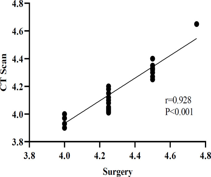
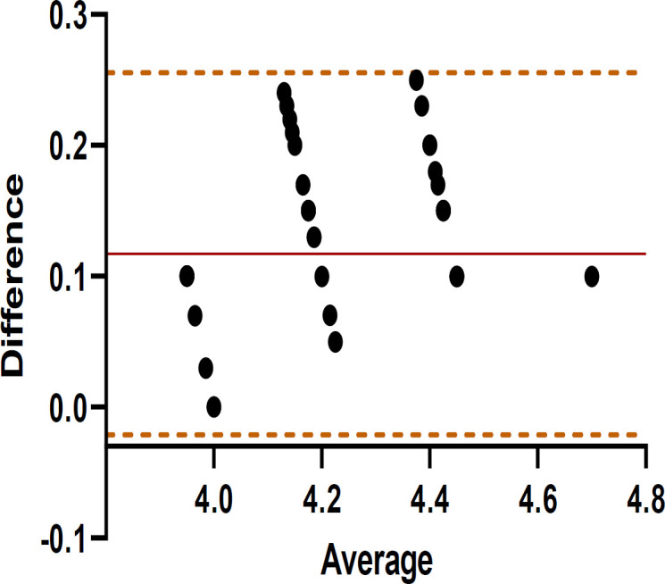
The mean distances measured by HRCT and intraoperatively were 4.15mm and 4.27mm, respectively. A strong and statistically significant correlation (r=0.928, P<0.001) was observed between the two approaches, indicating a robust association. The Bland-Altman analysis revealed a mean bias of 0.11±0.07mm, with limits of agreement (LoAs) ranging from -0.02 to 0.26 mm, and no points exceeding the 95% LoAs. The maximum potential error between the two measurement methods was 0.28mm, suggesting that HRCT imaging can reliably predict prosthesis length. In a stratified analysis based on the surgical distance (≤4 mm [N=11], 4.25mm [N=25], ≥4.5mm [N=13]), good agreement was maintained in the Bland-Altman analysis.
Preoperative HRCT imaging may be a valuable tool for accurately predicting the required prosthesis length prior to stapedotomy in otosclerosis patients.
本研究旨在评估术前高分辨率计算机断层扫描(HRCT)成像在测量砧骨长突至镫骨足板距离方面的准确性,以及其预测耳硬化症患者镫骨手术所需最佳假体长度的潜力。
本横断面研究纳入了50例计划进行初次镫骨手术的患者。一名放射科医生获取并重建了颞骨的术前HRCT扫描图像,以在轴向和冠状位视图中测量砧骨长突至椭圆窗的距离。然后将这些HRCT测量值与耳鼻喉科医生进行的术中测量值进行比较。使用相关性分析和布兰德-奥特曼分析评估两种方法之间的一致性。
HRCT测量的平均距离和术中测量的平均距离分别为4.15mm和4.27mm。两种方法之间观察到强且具有统计学意义的相关性(r = 0.928,P < 0.001),表明存在密切关联。布兰德-奥特曼分析显示平均偏差为0.11±0.07mm,一致性界限(LoA)范围为-0.02至0.26mm,且无点超出95%LoA。两种测量方法之间的最大潜在误差为0.28mm,表明HRCT成像能够可靠地预测假体长度。在基于手术距离(≤4mm [N = 11]、4.25mm [N = 25]、≥4.5mm [N = 13])的分层分析中,布兰德-奥特曼分析保持了良好的一致性。
术前HRCT成像可能是一种有价值的工具,可用于在耳硬化症患者进行镫骨手术前准确预测所需的假体长度。