Kanbe T, Tanaka K
Infect Immun. 1982 Nov;38(2):706-15. doi: 10.1128/iai.38.2.706-715.1982.
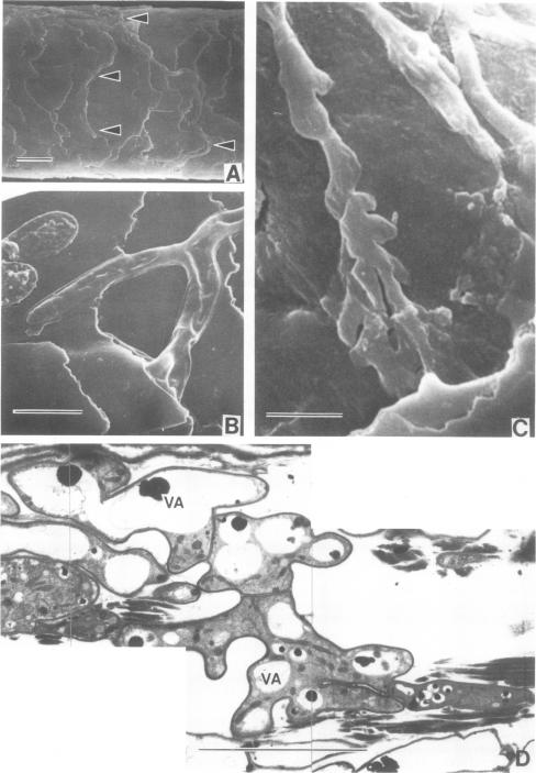
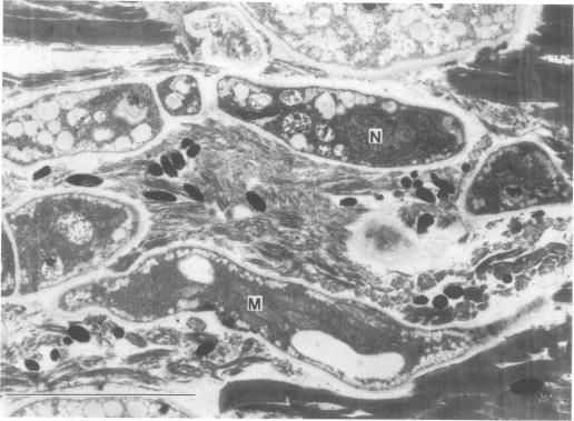
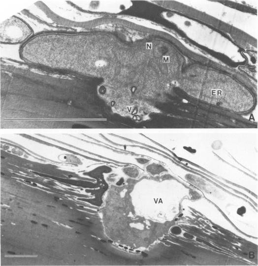
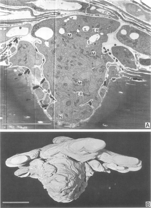
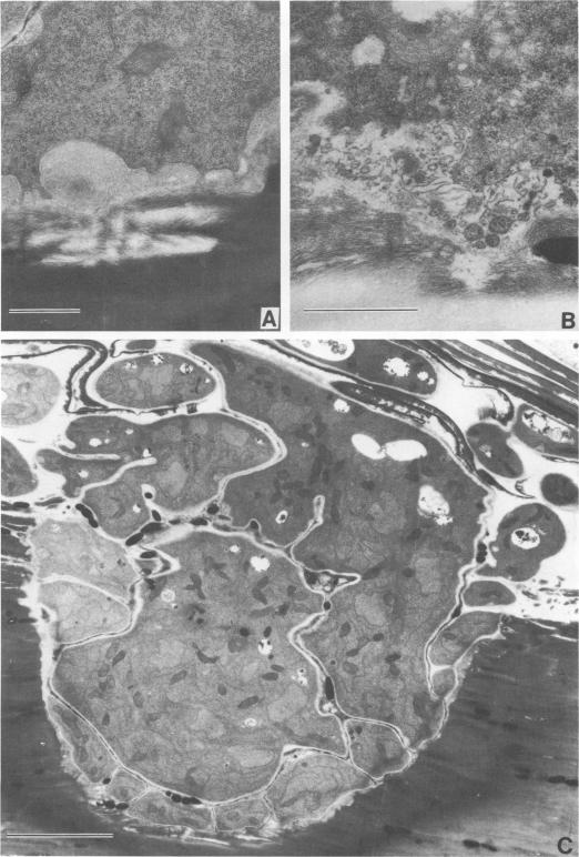
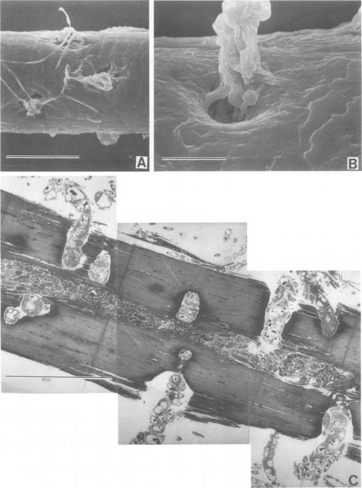
The pattern of invasion of human hair in vitro by the dermatophyte Microsporum gypseum was studied by transmission and scanning electron microscopy. Mycelia that invaded the hair cortex through the edge of cuticles showed a flattened "frond" growth in contrast to the filamentous form seen on ordinary laboratory media. The frond cells were characterized by the presence of vesicles formed by invaginations of plasmalemma, and lomasomes were prominent in the region adjacent to the hard keratinized tissue of the hair cortex being degraded as well. The initial perforating organ, which originated from the frond mycelium, appeared as an enlarged spherical cell which integrated with the laterally branched hyphae, as revealed by analysis of a three-dimensional model reconstructed from a series of sections. The fully developed perforating organ consisted of a column of wide and short cells which penetrated perpendicularly through the hair cortex. Through the medulla the filamentous hyphae had grown profusely in a longitudinal direction. Our studies confirm earlier light microscope observations and provide new ultrastructural details on the development of the eroding frond and the perforating organ.
通过透射电子显微镜和扫描电子显微镜研究了石膏样小孢子菌在体外对人毛发的侵袭模式。侵入毛发皮质的菌丝通过角质层边缘进入,与在普通实验室培养基上看到的丝状形态相比,呈现出扁平的“叶状体”生长。叶状体细胞的特征是存在由质膜内陷形成的囊泡,在与毛发皮质硬角质化组织相邻且正在降解的区域,边体也很突出。最初的穿孔器官起源于叶状体菌丝,表现为一个扩大的球形细胞,与横向分支的菌丝整合在一起,这是通过对一系列切片重建的三维模型分析揭示的。完全发育的穿孔器官由一列宽而短的细胞组成,这些细胞垂直穿透毛发皮质。通过髓质,丝状菌丝在纵向上大量生长。我们的研究证实了早期光学显微镜的观察结果,并提供了关于侵蚀叶状体和穿孔器官发育的新超微结构细节。