Narbaitz R, Bastani B, Galvin N J, Kapal V K, Levine D Z
Department of Anatomy and Neurobiology, University of Ottawa, Canada.
J Anat. 1995 Apr;186 ( Pt 2)(Pt 2):245-52.
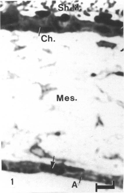
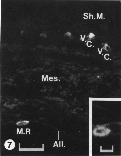
The chick embryo, confined in the eggshell, has to dispose/buffer the acid generated by its metabolism, as well as to release calcium from the shell which is used for growth. To localise H(+)-ATPase, electron microscope and immunocytochemical studies were conducted on chorioallantoic membranes of 15-17 d chick embryos. Ultrastructural studies of the villus cavity (VC) cells in the chorionic epithelium demonstrated that their apical plasma membrane, juxtaposed with the shell membranes, contains microvilli as well as microplicae which possess 9-10 nm studs at a density of 16,700 particles/micron2, a characteristic feature of the polarised H(+)-ATPase pump. Immunocytochemical staining, using a monoclonal antibody to the 31 kDa subunit of H(+)-ATPase, confirmed the presence of large amounts of the vacuolar H(+)-ATPase in the VC shells with a distribution highly polarised towards the eggshell membranes. Immunoelectron-microscopic localisation studies using a rabbit antiserum to whole bovine H(+)-ATPase and immunogold technique, confirmed the localisation of H(+)-ATPase at the apical microvilli/microplicae as well as in the subapical vesicles. In the allantoic epithelium, the presence of mitochondria-rich (MR) cells was confirmed; it was shown that these cells extend through the full thickness of this epithelium. The MR cells also contained large numbers of 9-10 nm studs, typical of proton secreting cells, in their apical plasma membrane. This was confirmed by immunocytochemical staining which showed abundant localisation of H(+)-ATPase in these cells; this localisation was, however, diffuse rather than apical.(ABSTRACT TRUNCATED AT 250 WORDS)
被限制在蛋壳内的鸡胚,必须处理/缓冲其新陈代谢产生的酸,同时还要从蛋壳中释放钙用于生长。为了定位H(+)-ATP酶,对15 - 17日龄鸡胚的绒毛尿囊膜进行了电子显微镜和免疫细胞化学研究。对绒毛膜上皮绒毛腔(VC)细胞的超微结构研究表明,其与壳膜并列的顶端质膜含有微绒毛以及微褶,微褶上有密度为16,700个颗粒/微米2的9 - 10纳米短棒,这是极化H(+)-ATP酶泵的特征性特征。使用针对H(+)-ATP酶31 kDa亚基的单克隆抗体进行免疫细胞化学染色,证实VC壳中存在大量液泡型H(+)-ATP酶,其分布高度极化于蛋壳膜。使用针对全牛H(+)-ATP酶的兔抗血清和免疫金技术进行免疫电子显微镜定位研究,证实H(+)-ATP酶定位于顶端微绒毛/微褶以及顶端下小泡中。在尿囊上皮中,证实存在富含线粒体(MR)的细胞;结果表明这些细胞贯穿该上皮的全层。MR细胞的顶端质膜中也含有大量9 - 10纳米短棒,这是质子分泌细胞的典型特征。免疫细胞化学染色证实了这一点,该染色显示这些细胞中H(+)-ATP酶大量定位;然而,这种定位是弥散的而非顶端定位。(摘要截断于250字)