Nukada H, McMorran P D
Department of Medicine, Univeristy of Otago Medical School, Dunedin, New Zealand.
J Anat. 1994 Oct;185 ( Pt 2)(Pt 2):259-66.
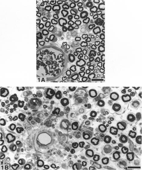
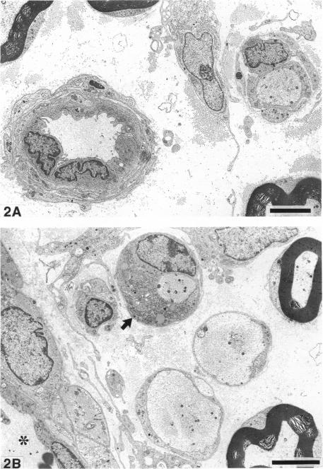
Nerve ischaemia plays a major role in the development of pathological alterations in various neuropathies, and the effects of ischaemia are amplified by reperfusion in various tissues. While pathological alterations in acutely ischaemic nerve have been established, nerve pathology resulting from reperfusion injury has never been elucidated. To evaluate what cell type in peripheral nerve is affected by reoxygenation following a hypoxic episode, we developed an animal model of transient severe limb ischaemia. Near-complete ischaemia, confirmed by the measurement of nerve blood flow, was achieved by clamping multiple arteries of supply to rat hindlimb. After 3, 5 or 7 h of limb ischaemia, vascular clips were released to reperfuse blood flow. Pathology in sciatic, tibial and peroneal nerves at the lower thigh level was examined at 7 d after reperfusion. All reperfused nerves developed demyelinated nerve fibres, particularly in perivascular regions. Although 3 h of ischaemia followed by reperfusion caused demyelination, perivascular demyelination was more prominent after a longer period of ischaemia with reperfusion. Two types of nerve oedema were observed; endoneurial oedema especially in perivascular and subperineurial spaces, and intramyelinic oedema. Nerve fibres with intramyelinic oedema were not confined to the perivascular region. Swollen endothelial cells in endoneurial vessels were also invariably observed. Nerve ischaemia per se, without reperfusion, did not induce these pathological changes. Because myelin appears to be particularly susceptible to activated free radicals, oxidative stress, activated neutrophils, and cytokine formation seem to be important underlying mechanisms in the development of perivascular demyelination and intramyelinic oedema in ischaemic/reperfused nerves.(ABSTRACT TRUNCATED AT 250 WORDS)
神经缺血在各种神经病变的病理改变发展过程中起主要作用,并且缺血的影响在各种组织中会因再灌注而放大。虽然急性缺血性神经的病理改变已得到确认,但再灌注损伤导致的神经病理变化从未被阐明。为了评估缺氧发作后外周神经中哪种细胞类型受复氧影响,我们建立了一个短暂性严重肢体缺血的动物模型。通过夹闭大鼠后肢的多条供血动脉实现了近乎完全的缺血,这通过测量神经血流得以证实。肢体缺血3、5或7小时后,松开血管夹以恢复血流。在再灌注7天后检查大腿下部水平的坐骨神经、胫神经和腓总神经的病理情况。所有再灌注的神经都出现了脱髓鞘神经纤维,特别是在血管周围区域。虽然缺血3小时后再灌注会导致脱髓鞘,但长时间缺血后再灌注时血管周围脱髓鞘更为明显。观察到两种类型的神经水肿:特别是在血管周围和神经束膜下间隙的神经内膜水肿,以及髓鞘内水肿。有髓鞘内水肿的神经纤维并不局限于血管周围区域。神经内膜血管中肿胀的内皮细胞也总是能被观察到。单纯的神经缺血,无再灌注,不会诱发这些病理变化。因为髓鞘似乎对活化的自由基特别敏感,氧化应激、活化的中性粒细胞和细胞因子形成似乎是缺血/再灌注神经中血管周围脱髓鞘和髓鞘内水肿发展的重要潜在机制。(摘要截短至250字)