Liu J Y, Morris G F, Lei W H, Corti M, Brody A R
Lung Biology Program, Tulane University School of Medicine, New Orleans, LA, USA.
Am J Pathol. 1996 Jul;149(1):205-17.
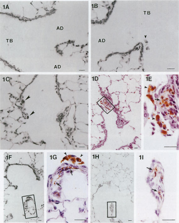
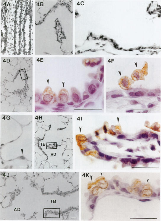
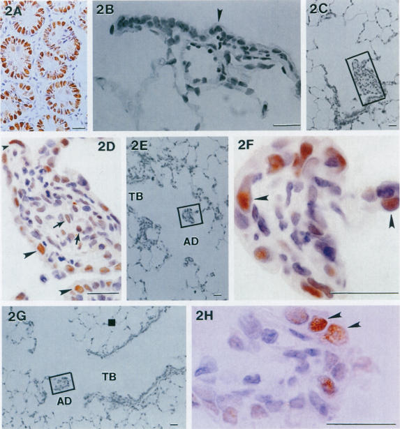
It has become apparent that the numerous growth factors and cytokines are produced during the development of fibroproliferative lung disease. Investigators must sort out which combinations of these factors are playing mechanistic roles in the disease process. Here we demonstrate that transforming growth factor (TGF)-alpha, a potent epithelial and mesenchymal cell mitogen, is upregulated specifically at the sites of asbestos fiber deposition in the lungs of rats exposed for 5 hours. Unexposed animals and those exposed to high concentrations of iron spheres exhibited no increase in TGF-alpha expression at any time during the experiment. Inhaled asbestos fibers deposit initially at the bronchiolar-alveolar duct regions and alveolar macrophages accumulate at these sites within hours. Non-isotopic in situ hybridization and immunohistochemistry were used to show that the mRNA that codes for TGF-alpha along with the peptide were clearly up-regulated at the bronchiolar-alveolar duct regions by 24 hours after the single asbestos exposure. The numbers of labeled cells demonstrated that expression of the mRNA and protein remained significantly above background for at least 2 weeks after exposure along with increased cell proliferation assessed by staining for proliferating cell nuclear antigen. This, to our knowledge, is the first demonstration of TGF-alpha expression at sites of lung injury in developing fibroproliferative disease. This finding supports the hypothesis that the growth factor is involved in the dramatic epithelial and mesenchymal proliferation we documented previously, although additional experiments will be essential to establish the precise role of TGF-alpha.
很明显,在纤维增生性肺病的发展过程中会产生多种生长因子和细胞因子。研究人员必须弄清楚这些因子的哪些组合在疾病进程中发挥着机制性作用。在此我们证明,转化生长因子(TGF)-α,一种强大的上皮和间充质细胞有丝分裂原,在暴露5小时的大鼠肺中石棉纤维沉积部位特异性上调。未暴露的动物以及暴露于高浓度铁球的动物在实验期间的任何时候TGF-α表达均未增加。吸入的石棉纤维最初沉积在细支气管-肺泡管区域,数小时内肺泡巨噬细胞在这些部位聚集。采用非同位素原位杂交和免疫组织化学方法显示,单次石棉暴露后24小时,编码TGF-α的mRNA以及肽在细支气管-肺泡管区域明显上调。标记细胞的数量表明,暴露后至少2周,mRNA和蛋白质的表达仍显著高于背景水平,同时通过增殖细胞核抗原染色评估的细胞增殖也增加。据我们所知,这是首次在发展中的纤维增生性疾病的肺损伤部位证明TGF-α的表达。这一发现支持了生长因子参与我们之前记录的显著上皮和间充质增殖的假说,尽管还需要进行更多实验来确定TGF-α的确切作用。