• 文献检索
  • 文档翻译
  • 深度研究
  • 学术资讯
  • Suppr Zotero 插件Zotero 插件
  • 邀请有礼
  • 套餐&价格
  • 历史记录
应用&插件
Suppr Zotero 插件Zotero 插件浏览器插件Mac 客户端Windows 客户端微信小程序
定价
高级版会员购买积分包购买API积分包
服务
文献检索文档翻译深度研究API 文档MCP 服务
关于我们
关于 Suppr公司介绍联系我们用户协议隐私条款
关注我们

Suppr 超能文献

核心技术专利:CN118964589B侵权必究
粤ICP备2023148730 号-1Suppr @ 2026

文献检索

告别复杂PubMed语法,用中文像聊天一样搜索,搜遍4000万医学文献。AI智能推荐,让科研检索更轻松。

立即免费搜索

文件翻译

保留排版,准确专业,支持PDF/Word/PPT等文件格式,支持 12+语言互译。

免费翻译文档

深度研究

AI帮你快速写综述,25分钟生成高质量综述,智能提取关键信息,辅助科研写作。

立即免费体验

白色中心性视网膜出血( Roth斑)

White-centred retinal haemorrhages (Roth spots).

作者信息

Ling R, James B

机构信息

Eye Unit, Stoke Mandeville Hospital, Aylesbury, Bucks, UK.

出版信息

Postgrad Med J. 1998 Oct;74(876):581-2. doi: 10.1136/pgmj.74.876.581.

DOI:10.1136/pgmj.74.876.581
PMID:10211348
原文链接:https://pmc.ncbi.nlm.nih.gov/articles/PMC2361020/
Abstract

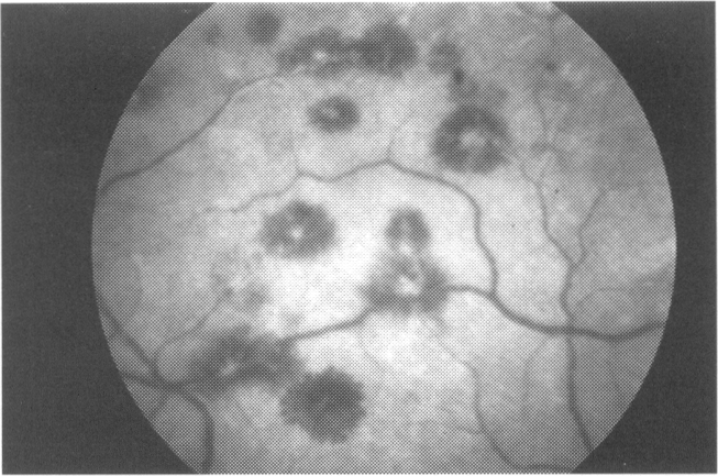
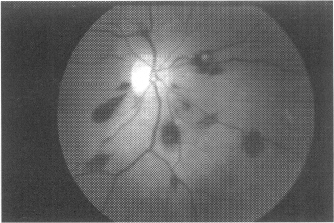
Roth spots (white-centred retinal haemorrhages) were classically described as septic emboli lodged in the retina of patients with subacute bacterial endocarditis. Indeed many have considered Roth spots pathognomonic for this condition. More recent histological evidence suggests, however, that they are not foci of bacterial abscess. Instead, they are nonspecific and may be found in many other diseases. A review of the histology and the pathogenesis of these white-centred haemorrhages will be provided, along with the work-up of the differential diagnosis.

摘要

Roth斑(中心白色的视网膜出血)传统上被描述为亚急性细菌性心内膜炎患者视网膜中滞留的脓毒性栓子。的确,许多人认为Roth斑是这种疾病的特征性表现。然而,最近的组织学证据表明,它们并非细菌性脓肿病灶。相反,它们是非特异性的,可能在许多其他疾病中出现。本文将对这些中心白色出血的组织学和发病机制进行综述,并介绍鉴别诊断的检查方法。

https://cdn.ncbi.nlm.nih.gov/pmc/blobs/7ef1/2361020/a7ffa99d62ca/postmedj00094-0008-b.jpg
https://cdn.ncbi.nlm.nih.gov/pmc/blobs/7ef1/2361020/aa6d6a71676e/postmedj00094-0008-a.jpg
https://cdn.ncbi.nlm.nih.gov/pmc/blobs/7ef1/2361020/a7ffa99d62ca/postmedj00094-0008-b.jpg
https://cdn.ncbi.nlm.nih.gov/pmc/blobs/7ef1/2361020/aa6d6a71676e/postmedj00094-0008-a.jpg
https://cdn.ncbi.nlm.nih.gov/pmc/blobs/7ef1/2361020/a7ffa99d62ca/postmedj00094-0008-b.jpg

相似文献

1
White-centred retinal haemorrhages (Roth spots).白色中心性视网膜出血( Roth斑)
Postgrad Med J. 1998 Oct;74(876):581-2. doi: 10.1136/pgmj.74.876.581.
2
Roth spots seen on ophthalmoscopy: diseases with which they may be associated.眼底镜检查发现的 Roth 斑:与之相关的疾病。
Conn Med. 1995 May;59(5):271-3.
3
Roth spots in pernicious anaemia.恶性贫血中的罗氏斑。
BMJ Case Rep. 2011 Apr 19;2011:bcr0120113734. doi: 10.1136/bcr.01.2011.3734.
4
Splinter haemorrhages, Osler's nodes, Janeway lesions and Roth spots: the peripheral stigmata of endocarditis.瘀点、奥氏结节、詹韦氏损害和罗氏斑:心内膜炎的外周体征。
Br J Hosp Med (Lond). 2013 Sep;74(9):C139-42.
5
Retinal findings in adult leukaemia: correlation with leukocytosis.成人白血病的视网膜表现:与白细胞增多的相关性。
Clin Lab Haematol. 1996 Jun;18(2):105-9. doi: 10.1046/j.1365-2257.1996.d01-217.x.
6
Clinicopathological correlation of retinal lesions. Subacute bacterial endocarditis.
Arch Ophthalmol. 1965 Nov;74(5):658-62. doi: 10.1001/archopht.1965.00970040660012.
7
Roth spots in native valve endocarditis.
J Am Osteopath Assoc. 2013 Nov;113(11):863. doi: 10.7556/jaoa.2013.063.
8
Images in clinical medicine. Roth spots in infective endocarditis.
N Engl J Med. 2014 Jun 19;370(25):e38. doi: 10.1056/NEJMicm1312093.
9
Drusen of the disc and retinal haemorrhages.视盘玻璃膜疣和视网膜出血。
Br J Ophthalmol. 1973 May;57(5):299-306. doi: 10.1136/bjo.57.5.299.
10
Images in cardiovascular medicine. Roth's spots.
Circulation. 1999 Mar 9;99(9):1271. doi: 10.1161/01.cir.99.9.1271.

引用本文的文献

1
Unilateral Roth spots secondary to uterine leiomyoma.子宫平滑肌瘤继发的单侧 Roth 斑。
BMJ Case Rep. 2025 Jul 18;18(7):e266496. doi: 10.1136/bcr-2025-266496.
2
Retinal Hemorrhages and Long-Term Ocular Outcomes in Neonatal Hypoxic-Ischemic Encephalopathy.新生儿缺氧缺血性脑病中的视网膜出血与长期眼部结局
Medicina (Kaunas). 2025 May 16;61(5):906. doi: 10.3390/medicina61050906.
3
Risk Factors for Neonatal White-Centered Retinal Hemorrhages: Clinical Implications for Prevention.新生儿以视盘为中心的视网膜出血的危险因素:预防的临床意义

本文引用的文献

1
White centered hemorrhages: their significance.白色中心出血:其意义
Ophthalmology. 1980 Jan;87(1):66-9. doi: 10.1016/s0161-6420(80)35277-9.
2
Hemorrhagic retinoschisis due to aplastic anemia.
Arch Ophthalmol. 1968 Oct;80(4):433-5. doi: 10.1001/archopht.1968.00980050435005.
3
White centered retinal hemorrhages in diabetic retinopathy.
Ophthalmology. 1987 Apr;94(4):388-92. doi: 10.1016/s0161-6420(87)33435-9.
4
Int J Gen Med. 2025 Mar 27;18:1747-1756. doi: 10.2147/IJGM.S505664. eCollection 2025.
4
Fundus Photography-Based Distribution of Retinal Hemorrhages in Newborns: Implications for Underlying Mechanisms.基于眼底摄影的新生儿视网膜出血分布:对潜在机制的启示
J Pers Med. 2025 Jan 19;15(1):38. doi: 10.3390/jpm15010038.
5
Retinal hemorrhages and damages from tractional forces associated with infantile abusive head trauma evaluated by wide-field fundus photography.广角眼底摄影评估与婴儿虐待性头部外伤相关的牵引性视网膜出血和损伤。
Sci Rep. 2024 Mar 4;14(1):5246. doi: 10.1038/s41598-024-54664-y.
6
Determinants for Anemic Retinopathy.贫血性视网膜病变的决定因素。
Beyoglu Eye J. 2023 May 1;8(2):97-103. doi: 10.14744/bej.2023.05658. eCollection 2023.
7
Eye: A window to the body.眼睛:身体的一扇窗户。
Indian J Ophthalmol. 2023 Jul;71(7):2648. doi: 10.4103/IJO.IJO_132_23.
8
Ophthalmic Manifestations as First Presenting Sign of Evans Syndrome.以眼部表现为首发症状的伊文氏综合征
J Vitreoretin Dis. 2022 Sep 13;6(6):479-484. doi: 10.1177/24741264211062931. eCollection 2022 Nov-Dec.
9
Roth-like Spot in an Adult With Vitamin B Deficiency and Nonepileptic Seizures.一名患有维生素B缺乏症和非癫痫性发作的成年人出现类罗氏斑。
J Vitreoretin Dis. 2021 Aug 13;6(5):412-413. doi: 10.1177/24741264211028788. eCollection 2022 Sep-Oct.
10
Clinical aspects of malarial retinopathy: a critical review.疟原虫性视网膜病变的临床方面:批判性评价。
Pathog Glob Health. 2023 Jul;117(5):450-461. doi: 10.1080/20477724.2022.2128568. Epub 2022 Oct 19.
Leukemic retinopathy. Relationship between fundus lesions and hematologic parameters at diagnosis.
白血病视网膜病变。诊断时眼底病变与血液学参数之间的关系。
Ophthalmology. 1989 Jun;96(6):860-4. doi: 10.1016/s0161-6420(89)32809-0.
5
Complications of glaucoma surgery. Ocular decompression retinopathy.
Arch Ophthalmol. 1992 Jul;110(7):965-8. doi: 10.1001/archopht.1992.01080190071032.