Anichini A, Molla A, Mortarini R, Tragni G, Bersani I, Di Nicola M, Gianni A M, Pilotti S, Dunbar R, Cerundolo V, Parmiani G
Department of Experimental Oncology Human Tumor Immunobiology Unit, Istituto Nazionale per lo Studio e la Cura dei Tumori, 20133 Milan, Italy.
J Exp Med. 1999 Sep 6;190(5):651-67. doi: 10.1084/jem.190.5.651.
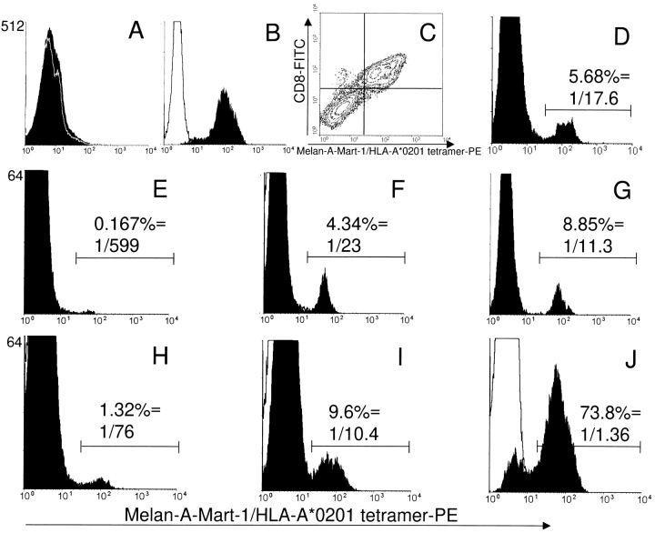
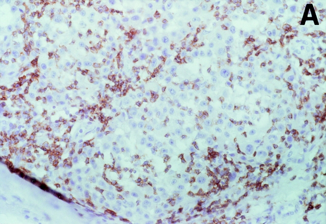
It is not known if immune response to T cell-defined human histocompatibility leukocyte antigen (HLA) class I-restricted melanoma antigens leads to an expanded peripheral pool of T cells in all patients, affects cytotoxic T lymphocyte (CTL) generation, and correlates with anti-tumor response in metastatic lesions. To this end, a limiting dilution analysis technique was developed that allowed us to evaluate the same frequency of peptide-specific T cells as by staining T cells with HLA-peptide tetrameric complexes. In four out of nine patients, Melan-A/Mart-1(27-35)-specific CTL precursors (CTLp) were >/=1/2,000 peripheral blood lymphocytes and found mostly or only in the CD45RO(+) memory T cell subset. In the remaining five patients, a low (<1/40,000) peptide-specific CTLp frequency was measured, and the precursors were only in the CD45RA(+) naive T cell subset. Evaluation of CTL effector frequency after bulk culture indicated that peptide-specific CTLs could be activated in all patients by using professional antigen-presenting cells as dendritic cells, but CTLp frequency determined the kinetics of generation of specificity and the final number of effectors as evaluated by both limiting dilution analysis and staining with HLA-A*0201-Melan-A/Mart-1 tetrameric complexes. Immunohistochemical analysis of 26 neoplastic lesions from the nine patients indicated absence of tumor regression in most instances, even in patients with an expanded peripheral T cell pool to Melan-A/Mart-1 and whose neoplastic lesions contained a high frequency of tetramer-positive Melan-A/Mart-1-specific T cells. Furthermore, frequent lack of a "brisk" or "nonbrisk" CD3(+)CD8(+) T cell infiltrate or reduced/absent Melan-A/Mart-1 expression in several lesions and lack of HLA class I antigens were found in some instances. Thus, expansion of peripheral immune repertoire to Melan-A/Mart-1 takes place in some metastatic patients and leads to enhanced CTL induction after antigen-presenting cell-mediated selection, but, in most metastatic lesions, it does not overcome tumor escape from immune surveillance.
尚不清楚针对T细胞定义的人类组织相容性白细胞抗原(HLA)I类限制性黑色素瘤抗原的免疫反应是否会在所有患者中导致外周T细胞池扩大,是否会影响细胞毒性T淋巴细胞(CTL)的生成,以及是否与转移性病变中的抗肿瘤反应相关。为此,开发了一种有限稀释分析技术,该技术使我们能够评估与用HLA-肽四聚体复合物对T细胞进行染色相同频率的肽特异性T细胞。在9名患者中的4名患者中,Melan-A/Mart-1(27-35)特异性CTL前体(CTLp)≥1/2000外周血淋巴细胞,且大多或仅存在于CD45RO(+)记忆T细胞亚群中。在其余5名患者中,测得的肽特异性CTLp频率较低(<1/40000),且前体仅存在于CD45RA(+)初始T细胞亚群中。大量培养后对CTL效应频率的评估表明,通过使用专业抗原呈递细胞(如树突状细胞),所有患者中的肽特异性CTL均可被激活,但CTLp频率决定了特异性生成的动力学以及效应细胞的最终数量,这通过有限稀释分析和用HLA-A*0201-Melan-A/Mart-1四聚体复合物染色来评估。对这9名患者的26个肿瘤病变进行的免疫组织化学分析表明,在大多数情况下不存在肿瘤消退,即使在那些外周T细胞池针对Melan-A/Mart-1扩大且其肿瘤病变中含有高频四聚体阳性Melan-A/Mart-1特异性T细胞的患者中也是如此。此外,在一些病变中经常缺乏“活跃”或“不活跃”的CD3(+)CD8(+)T细胞浸润或Melan-A/Mart-1表达降低/缺失,并且在某些情况下发现缺乏HLA I类抗原。因此,一些转移性患者的外周免疫库针对Melan-A/Mart-1发生了扩增,并在抗原呈递细胞介导的选择后导致CTL诱导增强,但在大多数转移性病变中,它并未克服肿瘤对免疫监视的逃逸。